INTRODUCTION
Nasal bone fractures are among the most commonly encountered types of pediatric facial bone fractures. The usual management of isolated nasal bone fractures is closed reduction to restore the appearance and function of the nose [1,2]. Although fracture reduction is simple, the postoperative results of the surgical correction of nasal bone fractures may be unsatisfactory [2-5]. Conservative treatment is performed in some cases if there are no external or functional problems because the fracture is linear with little displacement of the fracture segments, or upon the parent`s request. Bone union or bone remodeling can be expected to occur over time, but conservative treatment poses a risk of nasal deformity or related nasal complications [6,7]. The aim of this study was to evaluate the outcomes of bone remodeling based on changes in serial computed tomography (CT) examinations.
METHODS
A retrospective review was conducted of the medical records of patients under 12 years of age who received conservative treatment of a nasal bone fracture and underwent follow-up CT examinations upon parental request. The review identified 16 patients who were treated between March 2015 and December 2019. Information was extracted on patients’ demographic information (sex and age), follow-up period, the grade of the initial fracture according to the pattern of the fracture segments, and changes in the grade of the fracture over time.
Fracture evaluation was performed with a dual 128-channel CT scanner (SOMATOM Definition Flash; Siemens, Munich, Germany). We tried to use the same image slices of CT scans in order to minimize default error. Nasal bone fractures were classified using the system developed by Stranc and Robertson [8], as follows: plane I frontal impact (FI), plane II frontal impact (FII), plane I lateral impact (LI), plane II lateral impact (LII), and comminuted (C). On the initial and follow-up CT scans, the nasal bone fractures were graded using the following scores [9]: (1) excellent: no malalignment of the fracture segments is observed (i.e., without either a one-segment irregularity or displacement); (2) good: malalignment of the fracture segments is present (with either a one-segment irregularity or displacement); and (3) fair: malalignment of the fracture segments is present and both bony irregularity and displacement are observed for each segment (Table 1).
Bony contour improvement through bone remodeling was evaluated in terms of changes in the fracture grade between initial and subsequent CT scans. Four plastic surgeons evaluated the grade of the fracture, and the outcomes of bone remodeling was made by majority vote to ensure that the comparison was as objective as possible.
RESULTS
Of the 16 patients, 11 were male and five were female. The patients ranged in age from 2 to 11 years old, and the mean age was 6.2 years. The average follow-up period was 4.9 months (range, 25 days to 4 years). Five of the patients had FI fractures and 11 had LI fractures. On the initial CT images, in the FI group (n=5), the fractures were graded as good in three patients and fair in two patients. In the LI group (n=11), the fractures of nine patients were graded as good, and those of two patients were graded as fair. On the subsequent CT images, in the FI group (n=5), the fracture grade improved to excellent in three patients and to good in two patients. In the LI group (n=11), the fracture grade improved to excellent in nine patients and to good in one patient, while the remaining one patient showed no interval change from good to good (Table 2). Overall, bone remodeling showed favorable outcomes in 15 of 16 patients, as shown by improvements from the initial to subsequent CT scans from fair to excellent (n=1), fair to good (n=3) and good to excellent (n=11) (Table 3).
DISCUSSION
In pediatric patients with facial bone fractures, unless careful attention is paid to the initial diagnosis and management, the disturbance of the growth centers may cause posttraumatic deformities [1,2,10]. A particular issue is that nasal bone fractures in pediatric patients tend to be underestimated due to the underdevelopment of nasal bone, which is associated with relatively more extensive swelling and ecchymosis [10]. Furthermore, the parents of children with facial bone fractures often favor non-surgical interventions. For these reasons, conservative treatment is sometimes chosen in patients with mild nasal bone fractures without distinct displacement of the fracture segment. At some point after conservative treatment is chosen, parents may become worried about the nasal deformity or functional nasal problems, and therefore request an examination to assess the degree of improvement or recovery of the fracture, despite concerns about radiation exposure. X-ray imaging may not be a reliable diagnostic method for examining pediatric nasal bone fractures because nasal fractures that have not fully ossified may be inaccurately identified. Instead, facial bone CT is useful for more accurate evaluations of nasal bone fractures in children [11].
In our study, we reviewed the records of patients under 12 years of age who received conservative treatment of a nasal bone fracture and for whom follow-up CT examinations were performed upon parental request. All five patients in the FI group showed improvements in the bony contour of the fracture segments through remodeling (three from good to excellent and two from fair to good).
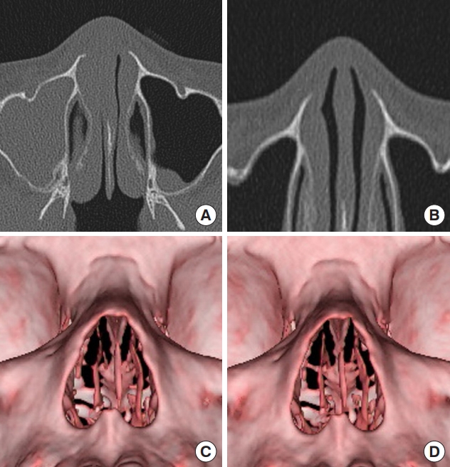
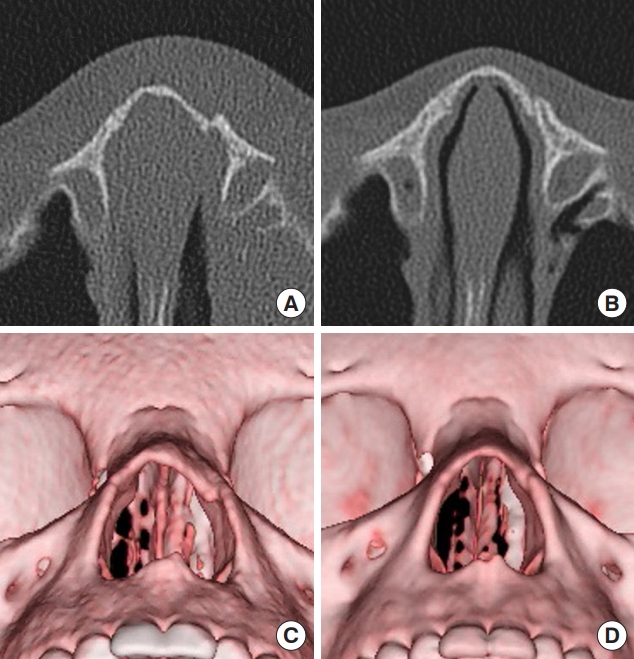
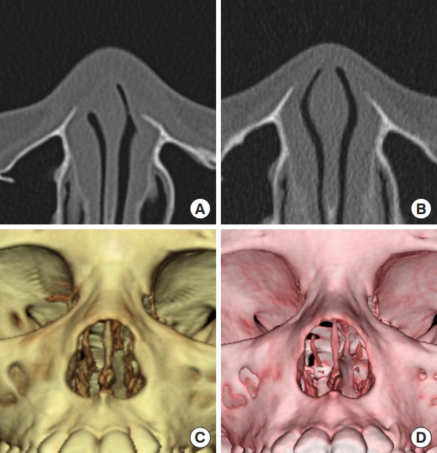
Fig. 1 presents an example of one of the three patients in whom the fracture was initially graded as good. In this patient, a fracture segment with a slight irregularity at the right lower end of the nasal bone and a non-excessively displaced fracture segment at the left lower end of the nasal bone were remodeled into a more favorable position over a follow-up of 45 days. Fig. 2 presents an example of one of the two patients in the FI group in whom the fracture was initially graded as fair. This patient had a greenstick fracture with displacement of one segment on the right nasal wall and an irregularity of the other segment on the left nasal wall. Over 70 days of follow-up, remodeling led to an improvement to a grade of good, with continuing displacement on the right nasal wall, but no irregularity of the other segment on the left nasal wall. Eight of the 11 patients in the LI group showed improvements from good to excellent, one patient from fair to excellent, and one patient from fair to good. In one patient, no interval change was shown. Of the nine patients with an LI fracture that was initially graded as good, eight had a linear fracture or little displacement of fracture segments. These patients improved from good to excellent over a mean follow-up period of 50.1 days. In contrast, one patient, who had a nasal wall fracture with wide displacement, did not show a serial change in fracture grade over a follow-up period of 25 days. Fig. 3 presents an example of a patient with an LI fracture that was initially graded as fair. This patient had an irregular nasal tip fracture and a left nasal wall fracture with displacement, but the parents refused surgical intervention. With conservative treatment, this patient’s fracture improved from fair to excellent over a long-term follow-up period of 19 months. Fig. 4 shows an example of a patient in the LI group with an initial fracture grade of good. This patient had a fracture segment with an irregularity on the frontal process of the left maxilla, and the fracture improved to excellent over a follow-up period of 42 days. After surgery for a nasal bone fracture, the nose usually maintains its pyramid, and deformities that occur as swelling decreases are traditionally considered to result from poor reduction [12].
Tremolet de Villers [13] noted that a nasal fracture segment reduced to an appropriate position may have a self-supporting force, resulting in favorable alignment of the nasal pyramid. The periosteum inside the nasal mucosa and skin and the connective tissues around the fracture are considered to be important factors for progressive bone remodeling as time passes and bony union occurs after reduction of a nasal bone fracture [14]. Likewise, if the fracture itself is mild, and if there is little displacement or minimal irregularity of the fracture segments, spontaneous improvements of the fracture segments into a favorable position through bone remodeling can be expected, even without surgical intervention. In this study, we identified bony contour improvements and alignment improvements of the nasal pyramid in both FI and LI fractures after conservative treatments through bone remodeling over the course of follow-up in pediatric patients with mild nasal bone fractures. In a single patient with a highly displaced or separated fracture segment, we could not identify interval changes of remodeling over the follow-up period of 25 days. However, we could see narrowing of displacement of fracture segment, and we think the result will be a little better after a more time.
The limitations of this study include the fact that the outcomes were not measured quantitatively, and our findings are difficult to generalize due to the small sample size. Nevertheless, these results suggest that if the displacement of fragment does not exceed the thickness of the nasal bone or if the end plate of an irregular fracture segment is continuous with unfractured bone, favorable bone remodeling of the fracture segments could be expected during long-term follow-up. Therefore, conservative treatment may be a feasible option for mild nasal fractures in pediatric patients.