Arch Craniofac Surg Search

CLOSE


Arch Craniofac Surg > Volume 23(4); 2022 > Article
Kim, Yang, and Oh: Correction of pixie ear: infraauricular skin redraping method

Abstract

Patients with pixie ear have an attached, tapering, and low-set earlobe. Traditional methods usually describe excision of the caudal portion of the lobule and reattachment in a more superior position. The present report suggests a simplified skin redraping method for correction of pixie ear. The procedure provides easy method to design and perform, which only requires elevation and trimming of the skin. Other ancillary procedures, such as flap design, anchoring, plication, and subdermal fixation, are not required. This method produces satisfactory results. Postoperative scar is invisible because the incision is on the retroauricular region, and the corrected earlobe has a more natural appearance than the repositioned earlobe. Moreover, skin redraping avoids tension, which contributes to minimization of the postoperative scar.

INTRODUCTION

Pixie ear is a noticeable deformity [1] that appears as an attached, tapered, and low-set earlobe [2]. It can be congenital or acquired, and acquired causes include trauma, rhytidectomy, or total reconstruction of the ear [1,3,4]. Traditional methods have been reported, but they usually excise the lobule and reattach it in a superior position [5,6]. The scar along the border of the earlobe, however, disturbs the ear’s natural appearance and can become hypertrophied [6]. In addition, the surgery is combined with fat or dermofat graft in the earlobe [7] and performed with complicated plication suture around the earlobe [2].
The present report suggests a simple skin redraping method for correction of pixie ear without a visible scar. This concept is modified and based on medial epicanthoplasty using the skin redraping method [8]. Like skin redraping epicanthoplasty removes the hood of the epicanthal fold and expands the medial canthus, skin redraping correction of pixie ear removes the hood of the pixie earlobe and forms an intertragal notch between the earlobe and the jaw line. Furthermore, the scar sits in the retroauricular area and is inconspicuous when viewed from the front.

IDEA

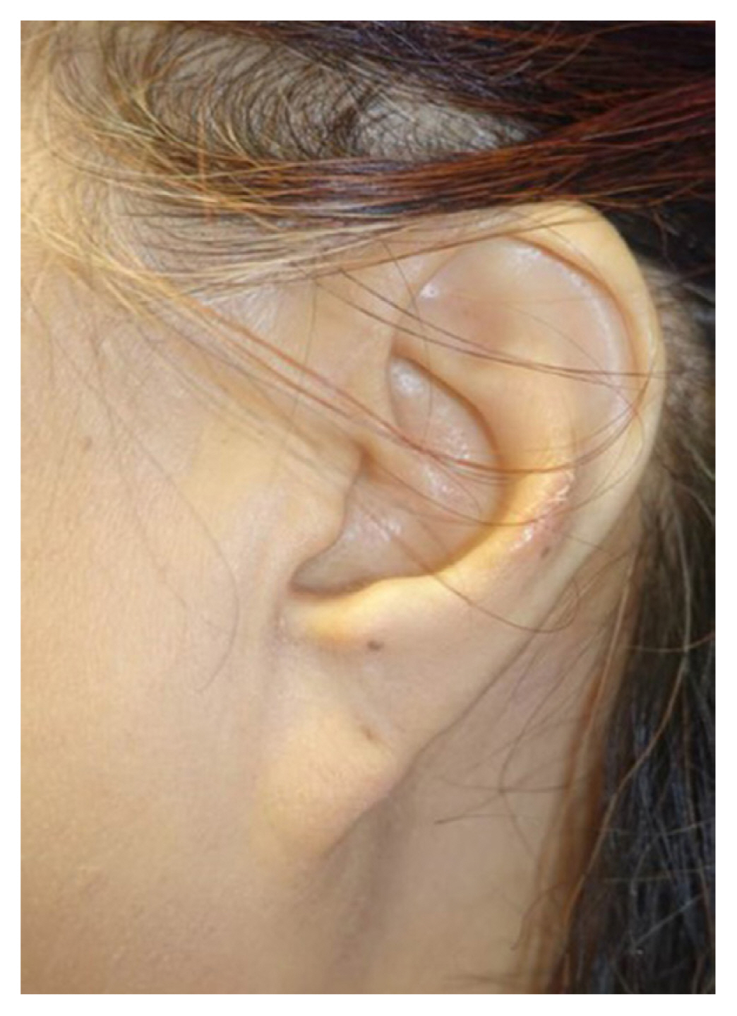
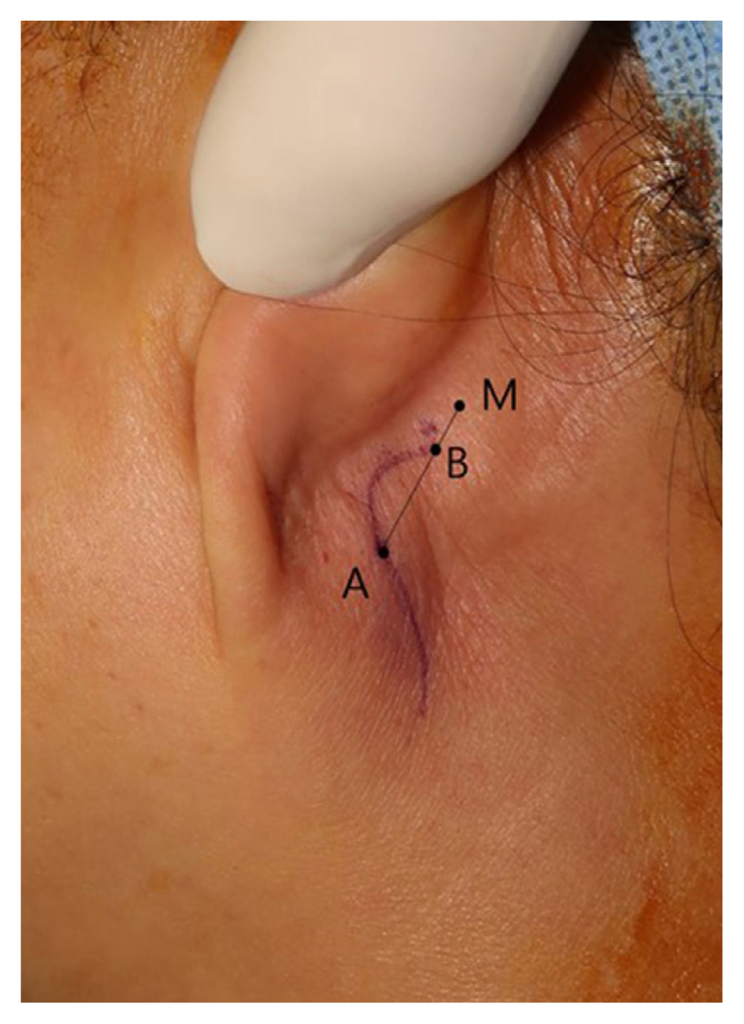
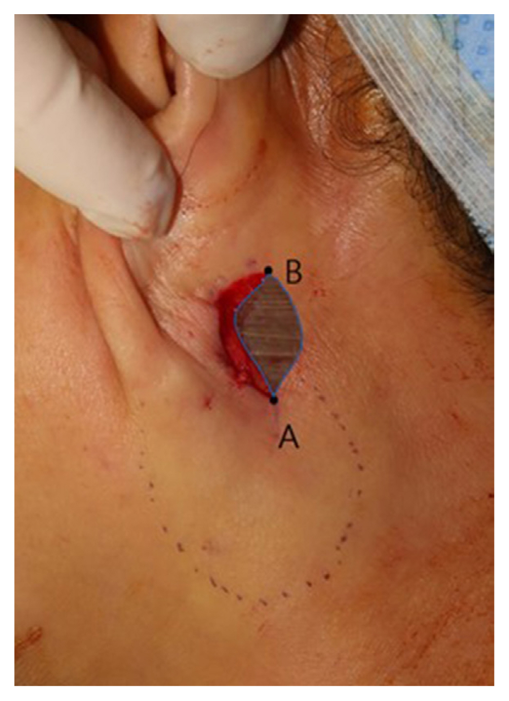
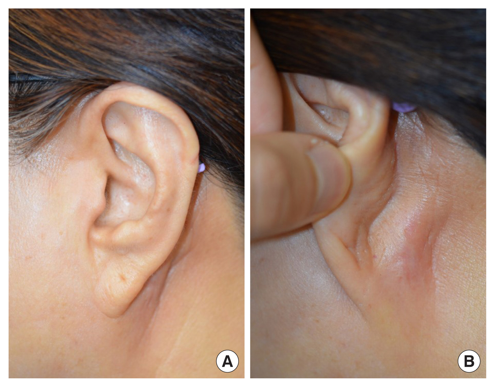
The present method is simple to design, easy to perform, and can be performed under local anesthesia. We observed a case of congenital pixie ears with tapered and low-set earlobes (Fig. 1). The patient was a 64-year-old woman who had congenital bilateral pixie ears and was dissatisfied with her appearance. Pixie ear was hereditary, and her two sisters also had it. She has always wanted to take correction on it. The procedure required only elevation of the skin and trimming of the redundant skin. The incision was planned in the retroauricular region; we took the position 1 cm posterior to the midpoint of the earlobe as point A, and the center of the mastoid process as point M. In the line connecting points A and M, point B, which is close to point M by 2:8, was determined as the suture point because of tension on around skin (Fig. 2). After the skin was incised along the design, the neck skin on infraauricular area was undermined in subcutaneous layer and elevated for redraping. Point A was approximated toward point B and sutured without tension. The remnant skin between point A and point B was trimmed (Fig. 3). The patient reported a satisfactory result after 3 years of surgery (Fig. 4).

DISCUSSION

The skin redraping method for correction of pixie ear is simple and easy to perform. The corrected earlobe shows a more natural appearance than the earlobe repositioned through conventional surgery. The scar appears only in the retroauricular region, so it is unnoticeable. Skin redraping avoids tension, which is another important factor contributing to scar minimization.
Pixie ear is a noticeable deformity [1], but it has not received as much attention as microtia, anotia, prominent ear, or traumatic ear [2,9,10]. Pixie ear has an attached, tapered, and low-set earlobe and can be congenital or acquired [14]. Acquired factors include trauma, rhytidectomy, and total reconstruction of ear, among which rhytidectomy results in excessive skin resection around the ear or insufficient suspension of the deep soft tissues [1,11].
Traditional methods for pixie ear have been described. Most of the methods excise the caudal portion of the lobule and reattach it in a more superior position [5,6]. Such methods, however, result in smaller earlobes [7], or the scar along the border of the earlobe disturbs the ear’s natural appearance and can become hypertrophied [6]. In addition, correction of pixie earlobe is combined with dermofat graft to increase the volume of the earlobe [7].
The concept of the present method is modified from medial epicanthoplasty using the skin redraping method [8]. Skin redraping epicanthoplasty removes the hood of the epicanthal fold and widens the medial canthus. Likewise, skin redraping correction of pixie ear removes the hood of the pixie earlobe and shapes the intertragal notch between the earlobe and the jaw line.
In this method, the scar sits in the retroauricular area and is inconspicuous when viewed from the front. Finally, skin redraping for pixie ear creates a natural appearance with tension-free and invisible scars in the retroauricular region.
The skin redraping method for correction of pixie ear is simple and easy and creates a natural appearance with tension-free and invisible scars in the retroauricular region.

Notes

Conflict of interest

No potential conflict of interest relevant to this article was reported.

Ethical approval

The study was approved by the Institutional Review Board of The Catholic Medical Center Office of Human Research Protection Program (IRB No. KC22ZISI0222).

Patient consent

The patient provided written informed consent for the publication and the use of her images.

Author contribution

Conceptualization: Jun Hyeok Kim, Deuk Young Oh. Data curation: Young Bin Yang. Visualization: Jun Hyeok Kim, Young Bin Yang. Writing - original draft: Jun Hyeok Kim. Writing - review & editing: Deuk Young Oh. Resources: Young Bin Yang. Supervision: Deuk Young Oh.

Fig. 1
Preoperative photograph of pixie ear.
acfs-2022-00675f1.jpg
Fig. 2
Design of the incision in the retroauricular region. Point A: 1 cm posterior to the midpoint of the earlobe; point M: the center of the mastoid process; and point B: close to point M by 2:8 in the line connecting points A and M.
acfs-2022-00675f2.jpg
Fig. 3
Intraoperative photograph. The dotted round line indicates the area of skin elevation for redraping. Point A is approximated to point B and sutured without tension. The remnant skin indicated by the shading area is trimmed.
acfs-2022-00675f3.jpg
Fig. 4
Three-year follow-up photographs. (A, B) Corrected and natural earlobe without noticeable scarring.
acfs-2022-00675f4.jpg

REFERENCES

1. Fuente-Del-Campo A. Commentary on: an update on the posterior earlobe/concha mastoid suspension suture to correct “pixie ear” following facelift: review of 40 cases. Aesthet Surg J 2021;41:NP1366-7.
crossref pmid pdf
2. Hoefflin SM. Simple repair of a pixie earlobe. Plast Reconstr Surg 2001;107:1623-4.
crossref pmid
3. Clevens RA, Baker SR. Plastic and reconstructive surgery of the earlobe. Facial Plast Surg 1995;11:301-9.
crossref pmid
4. Nakamura M, Ikeda T, Shioya N. Side-to-side earlobe variations with respect to surface area and shape: a quantitative study. Aesthetic Plast Surg 1995;19:561-5.
crossref pmid pdf
5. Rich JD, Gottlieb V, Shesol BF. A simple method for correction of the pixie earlobe. Plast Reconstr Surg 1982;69:136-8.
crossref pmid
6. Khoo BC. The pixie earlobe: a method of correction. Plast Reconstr Surg 1985;76:636-8.
crossref pmid
7. Park C. A new method for the correction of small pixie earlobe deformities. Ann Plast Surg 2007;59:273-6.
crossref pmid
8. Oh YW, Seul CH, Yoo WM. Medial epicanthoplasty using the skin redraping method. Plast Reconstr Surg 2007;119:703-10.
crossref pmid
9. Hwang K. White radish and swine scapular cartilage models for auricular framework carving training. Arch Craniofac Surg 2020;21:225-8.
crossref pmid pmc pdf
10. Lee D, Kim YS, Roh TS, Yun IS. Cryptotia recurrence lowering technique with additional acellular dermal matrix graft. Arch Craniofac Surg 2019;20:170-5.
crossref pmid pmc pdf
11. Kim DG, Cho HG, Ryu JY, Lee JS, Lee SJ, Lee JM, et al. Clinical assessment and management of auricular arteriovenous malformation: retrospective study. Arch Craniofac Surg 2021;22:141-7.
crossref pmid pmc pdf
TOOLS
Share :
Facebook Twitter Linked In Google+ Line it
METRICS Graph View
  • 1 Crossref
  •   Scopus
  • 2,693 View
  • 93 Download
Related articles in ACFS


ABOUT
ARTICLE CATEGORY

Browse all articles >

BROWSE ARTICLES
AUTHOR INFORMATION
Editorial Office
Dept. of Plastic and Reconstructive Surgery Chonnam National University Medical School, 42 Jebong-ro, Dong-gu, Gwangju 61469, Korea
Tel: +82-62-220-6354    Fax: +82-62-220-6357    E-mail: office_acfs@kcpca.or.kr                

Copyright © 2024 by Korean Cleft Palate-Craniofacial Association.

Developed in M2PI

Close layer
prev next