Arch Craniofac Surg Search

CLOSE


Arch Craniofac Surg > Volume 23(6); 2022 > Article
Kim and Kim: Current concepts of Kimura disease: pathophysiology and evolution of treatment

Abstract

Kimura disease (KD) is a chronic inflammatory disorder that frequently involves the subcutaneous tissue of the head and neck regions. It often manifests as regional lymphadenopathy or salivary gland enlargement in the affected area. The histologic architecture of the lymph nodes in KD patients is preserved, while the affected tissues show follicular hyperplasia and eosinophilic infiltration. No single modality has been adopted as an optimal treatment for KD. This article concentrates on the fundamental features of KD and reviews current approaches to its treatment.

Abbreviations:

ALHE,

angiolymphoid hyperplasia with eosinophilia
;

IgE,

immunoglobulin E
;

IL,

interleukin
;

KD,

Kimura disease
;

PTK,

protein tyrosine kinase
;

Th2,

T-helper 2
.

INTRODUCTION

Kimura disease (KD) is a rare chronic inflammatory condition of unknown etiology that usually presents as soft tissue tumors of the head and neck, especially in the retroauricular area, scalp, and forehead of young to middle-aged Asian men. It is often accompanied by regional lymphadenopathy, elevated serum immunoglobulin E (IgE) levels, and raised serum eosinophil counts [14]. The histologic findings include proliferating blood vessels and eosinophilic infiltration, which suggest an inflammatory response associated with an allergic or an autoimmune reaction to an unknown stimulus [5]. Surgery enables the greatest diagnostic accuracy and has been the mainstay of treatment. However, the optimal treatment of KD remains a matter of debate, and other options besides surgery have emerged [1,5]. This article reviews the fundamental features of KD and current knowledge regarding its treatment.

HISTORY

KD was first described by Kim in 1937 [6] as an “eosinophilic hyperplastic lymphogranuloma”. A decade later, a systemic description that included the clinical entity of unusual granulation combined with hyperplastic changes of lymphatic tissues in a self-limited allergic or autoimmune process was established by Kimura [7], for whom the disease was named. Although the condition is prevalent in East Asia in an endemic form, relatively few cases have been reported in the Western literature, in African Americans and Caucasians as well as individuals of Arab ancestry [4,810].

HISTOPATHOLOGY

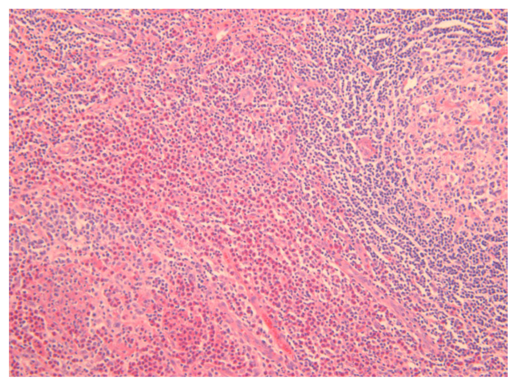
The histologic features of KD include follicular hyperplasia with reactive germinal centers; eosinophilic infiltrates involving the inner follicular areas, sinusoidal areas, perinodal soft tissues, and subcutaneous tissues; and the proliferation of postcapillary venules while preserving the nodal architecture (Fig. 1) [5,1115]. Chen et al. [11] reviewed 21 cases of KD and observed the following histologic features; eosinophils, necrosis, proteinaceous deposits and vascularization of germinal centers, polykaryocytes, eosinophilic folliculolysis, eosinophilic micro-abscesses, stromal sclerosis, perivenular sclerosis, rare giant cells, or small eosinophilic granulomas [5,11,16]. Various degrees of sclerosis are found in KD, and immunohistochemical staining for IgE reveals a reticular pattern of germinal centers [11].

PATHOGENESIS

The etiology and pathogenesis of KD are still unknown despite numerous studies. Although increased levels of IgE, tumor necrosis factor α, interleukin (IL)-4, IL-5, IL-13 and mast cells in peripheral blood, as well as in the affected lesions with eosinophilia, have been observed in most patients, no specific antigens have been identified [1719]. Allergic reactions, infections, neoplasms, and autoimmune reactions with an aberrant immune reaction have been suggested as possible etiologies [2022]. However, as Lu et al. [19] reported in a single case of KD secondary to renal allograft failure involving chronic graft rejection induced by T-helper produced ILs, T-helper 2 (Th2) cytokines, overgrowth of CD4+ (cluster of differentiation 4) cells, and Th2 may play an important role in KD. Furthermore, molecular studies of the immunoglobulin heavy chain and T-cell receptor gene support this reactive process. Clonal T-cell rearrangement was also described in a case report by Chim et al. [23] and confirmed by complete sequencing of the VDJ rearrangement [5,23,24].

CLINICAL FEATURES

Most lesions in KD involve deep subcutaneous tissues of the head and neck area, presenting as one or several deeper masses. The masses are observed as non-tender and poorly circumscribed with pruritus and pigment deposition in rare cases [1,25]. Commonly involved regions include the periauricular area, epicranium, orbit, and eyelids, while other sites such as the groin, extremities, and the soft tissue of the trunk are less often affected. Rare cases have been reported involving the hard palate, the larynx, and the median nerve [9,2527]. The majority of these masses frequently involve regional lymphadenopathy or salivary gland enlargement. However, most patients do not suffer from systemic symptoms, such as fever, night sweats, and weight loss. Coexisting renal disease from glomerulonephritis can be manifested by concomitant proteinuria and present as nephrotic syndrome with increased serum creatinine [12,28, 29]. Renal impairment might originate from immunocomplex-mediated damage or Th2 immune response disorders [28]. These renal diseases usually occur simultaneously with skin lesions [30].

DIAGNOSIS

Since there is no definite single specific diagnostic tool for KD, biopsy or excision of the involved mass or lymph node is frequently required for a pathological diagnosis. The histopathological findings of KD are similar, regardless of the anatomical site of involvement, and are characterized by a marked reactive follicular hyperplasia surrounded by a large number of eosinophils, lymphocytes and mast cells, sometimes forming micro-abscesses with vascular proliferation [16,31]. The vessels maintain thin walls of cuboidal endothelial cells, and the polykaryocytic Warthin-Finkeldey type of giant cells is a common finding. These findings are also present in lymph node biopsies, which show lymphoproliferation with prominent lymph follicle hyperplasia and germinal center enlargement while the nodal architecture is preserved [32,33].
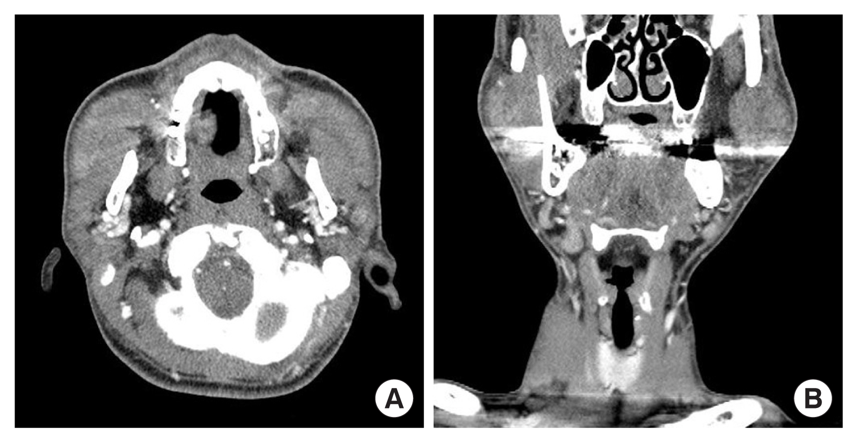
The radiologic findings of masses may aid in diagnosis of KD and differentiate them from other soft tissue tumors. On computed tomography with contrast enhancement, ill-defined enlarged subcutaneous masses with homogeneous, hyperattenuated lymph nodes and swollen salivary glands appear, reflecting the vascular nature of the lesions (Fig. 2) [29,34,35]. On magnetic resonance imaging, the lesions show heterogeneity, with both hypointense and mixed or high signal intensity on T1-weighted images and hyperintense signals on T2-weighted images. The degree of enhancement is variable and relates to the degree of fibrosis and vascular hyperplasia [35,36]. On positron emission tomography-computed tomography, KD may resemble neoplastic disorders, such as lymphoma or metastatic lymphadenopathy [37].
The differential diagnosis includes angiolymphoid hyperplasia with eosinophilia (ALHE), angioimmunoblastic T-cell lymphoma, Hodgkin lymphoma, Langerhans cell histiocytosis, Castleman disease, florid follicular hyperplasia, dermatopathic lymphadenopathy, allergic granulomatosis of Churg and Strauss, drug reaction, tumor node metastases, and parasitic lymphadenitis. KD can be confused with ALHE, which was often considered identical to KD in the past literature. However, ALHE is considered to be a type of endothelial neoplasm related to inflammatory stimulation, while KD is a chronic inflammatory disorder. ALHE often affects middle-aged Western women presenting with a well-circumscribed subcutaneous mass in the head and neck region, normally without regional lymphadenopathy, serum eosinophilia, or elevated IgE levels. Microscopically, ALHE shows the vascular endothelium forming aggregates and lobules lined by plump cuboidal or hobnail endothelial cells, which frequently involve large muscular vessels (Table 1) [7,17,38,39].

EVOLUTION OF TREATMENT

Several interventions have been reported in the previous literature; however, there is a lack of evidence on the optimal treatment for KD. Furthermore, owing to the rarity of KD, most clinical studies have been case reports or series reports; therefore, debate continues regarding the optimal treatment of KD.

Corticosteroids

Previous studies have reported the efficacy of systemic steroid therapy. Steroids are used to control local lesions, lymphadenopathy, and nephrotic syndrome. In particular, a patient with nephrotic syndrome was responsive to high-dose systemic steroids, with disappearance of proteinuria 7 months after treatment [28]. Another case report from China also revealed that the majority of patients included in the study responded well to treatment with corticosteroids alone [40]. However, Nakahara et al. [41] reported that local recurrence was frequently observed during steroid dose tapering or drug withdrawal and stated that long-term steroid use may cause digestive ulcers, osteoporosis, and acquired diabetes mellitus [4143]. Therefore, it is reasonable to use a steroid as a second-line treatment in KD while considering the risk of recurrence and side effects.

Immunosuppressive medications

According to a few recent studies, cyclosporine A is effective for recurrent KD patients [17,41,4448]. According to these reports, cyclosporine A was usually used for combined therapy with steroids to induce re-remission after KD relapse [49]. Cyclosporine A inhibits calcineurin, which signals IL-2 gene transcription in lymphocytes. The reduction of IL-2 inhibits T cell proliferation and suppresses T cell-mediated immune responses [17,50,51]. Moreover, Katagiri et al. [17] reported that cyclosporine A also decreased mRNA levels of IL-4, IL-5, and IL-13, resulting in the presence of fewer peripheral eosinophils and lower serum levels of IgE.
Omalizumab is an anti-IgE monoclonal antibody that binds to free IgE in the blood, which in turn, inhibits the activation of inflammatory mechanisms. As KD is suspected to be an IgE-mediated allergic disorder, a single pilot study reported evidence of omalizumab for treating KD. According to that study, the size of the masses and eosinophil counts of peripheral blood all decreased after omalizumab administration [52]. Moreover, omalizumab was also reported to be available for patients whose total serum IgE levels were below <1,500 kU/L in another case report. Therefore, omalizumab, as well as anti-IgE therapy, might have the potential to become definite treatment option for KD [53].
Imatinib, an inhibitor of protein tyrosine kinase (PTK), selectively blocks PTK and works effectively for steroid-resistant patients in hypereosinophilic syndrome. Since the tissues of KD patients are positive for expression of PTK with hypereosinophilic conditions, the authors of a previous study inferred that imatinib might be an effective drug for KD. However, further studies with actual clinical data are required for imatinib to be considered as a treatment option for KD [1,54].

Radiotherapy

Radiotherapy is considered an appropriate option for recurrent cases or poor surgical candidates. Hareyama et al. [55] reported that a 90% local control rate was achieved by using 26–30 Gy for irradiation and stated that the radiation field should be confined to the lesion and regional lymph nodes [55,56]. However, other reports showed that surgical excision combined with postoperative radiotherapy achieved a much lower local recurrence rate than surgery or radiotherapy alone [57]. Therefore, postoperative radiotherapy may be effective in controlling the residual lesion, reducing the recurrence rate without notable side effects.

Surgical excision

Surgery may allow the greatest diagnostic accuracy and has been the mainstay of treatment. However, as KD tends to be ill-defined on pathological examinations, it is difficult to achieve negative margins during surgical excision, which leads to a relatively high (25%) reported recurrence rate; furthermore, it was revealed that positive margins were a risk factor for disease recurrence [42,58,59]. Therefore, in clinical practice, surgical resection combined with low-dose postoperative radiotherapy has become the main treatment modality for KD.

Other interventions

In addition to conventional therapies, many other treatment options have been suggested. Suplatast tosilate, an anti-allergy drug, attenuated hyper-IgE and eosinophilia, suggesting that other mechanisms than the T cell-mediated immune response are related to the pathogenesis of KD. Cetirizine, which is another antiallergic agent, also showed beneficial effects in KD patients [60,61]. Boulanger et al. [62] reported that all-trans retinoic acids in combination with steroid therapy led to a rapid disappearance of clinical symptoms of KD. It was postulated that all-trans retinoic acids might exert immunomodulatory effects on Th2 cytokines and inhibit IL-4-mediated IgE production, causing the symptoms to disappear. A case report stated that sleep apnea due to KD improved with combination therapy of laser surgery, a steroid, and oral administration of pranlukast, which is a leukotriene receptor blocker [26]. Chewable nicotine also showed a treatment effect in skin disorders with eosinophilic infiltration [63]. A pediatric patient who had poor adherence to oral medications was treated with intravenous vincristine, resulting in remission of nephrotic syndrome complicated by KD and submandibular swelling [64]. However, these unconventional treatment options should be supported by large-scale clinical evidence, which requires further research.

PROGNOSIS

Even though a high overall recurrence rate (25%) has been reported with current treatment options–surgical excision (30.5%), medication (45%), and radiotherapy (60%)–KD has a favorable prognosis with no potential for malignancy. Sakamoto et al. [14] found that the eosinophil count was related to the therapeutic response in patients with KD and suggested that the eosinophil count might be used as a predictive marker for KD. According to other studies, patients who developed recurrence after surgical excision tended to have a maximal tumor diameter ≥3 cm, disease duration ≥5 years, peripheral eosinophil count ≥20%, or a serum IgE level ≥10,000 IU/mL, which required adjuvant therapy combined with surgical excision [2,57].

CONCLUSION

KD is a rare chronic inflammatory disorder with unknown etiology that frequently involves the subcutaneous tissue of the head and neck region. It is usually accompanied by regional lymphadenopathy and salivary gland involvement in Asian males. The distinct histologic features of KD include follicular hyperplasia with eosinophilic infiltration and preserved nodal architecture, making the diagnosis clear. Surgical resection combined with low-dose postoperative radiotherapy achieves the lowest local recurrence rate, and additional studies of immunosuppressive medications are required.

Notes

Conflict of interest

Han Koo Kim is an editorial board member of the journal but was not involved in the peer reviewer selection, evaluation, or decision process of this article. No other potential conflicts of interest relevant to this article were reported.

Author contributions

Conceptualization: Han Koo Kim. Formal analysis: Woo Ju Kim, Han Koo Kim. Visualization: Woo Ju Kim. Writing - original draft: Woo Ju Kim. Writing - review & editing: Han Koo Kim.

Fig. 1
Histopathological image of a cheek mass from a 33-year-old man with Kimura disease. Lymphoid hyperplasia with massive eosinophil infiltration (hematoxylin and eosin stain, ×200).
acfs-2022-01053f1.jpg
Fig. 2
Enhanced computed tomography shows ill-defined enlarged subcutaneous masses with hyperattenuated lymph nodes. (A) Axial view. (B) Coronal view.
acfs-2022-01053f2.jpg
Table 1
Clinical differences between Kimura disease and ALHE
Kimura disease ALHE
Clinical features Usually, solitary subcutaneous lesions with regional lymphadenopathy and salivary gland involvement
Predominant in Asian males
Small, superficial dermal erythematous nodules accompany pruritus, and bleeding
Occurs in all racial groups, with a female predominance
Histologic findings Marked reactive follicular hyperplasia with prominent follicles
Increased eosinophils, lymphocytes, and mast cells
Occasional formation of micro-abscesses with vascular proliferation
Vascular proliferation
Endothelium forming aggregates and lobules lined by plump cuboidal or hobnail endothelial cells

ALHE, angiolymphoid hyperplasia with eosinophilia.

REFERENCES

1. Sun QF, Xu DZ, Pan SH, Ding JG, Xue ZQ, Miao CS, et al. Kimura disease: review of the literature. Intern Med J 2008;38:668-72.
crossref pmid
2. Lee CC, Feng IJ, Chen YT, Weng SF, Chan LP, Lai CS, et al. Treatment algorithm for Kimura’s disease: a systematic review and meta-analysis of treatment modalities and prognostic predictors. Int J Surg 2022;100:106591.
crossref pmid
3. Yuen HW, Goh YH, Low WK, Lim-Tan SK. Kimura’s disease: a diagnostic and therapeutic challenge. Singapore Med J 2005;46:179-83.
pmid
4. Hobeika CM, Mohammed TL, Johnson GL, Hansen K. Kimura’s disease: case report and review of the literature. J Thorac Imaging 2005;20:298-300.
pmid
5. Abuel-Haija M, Hurford MT. Kimura disease. Arch Pathol Lab Med 2007;131:650-1.
crossref pmid pdf
6. Kim HT. Eosinophilic hyperplastic lymphogranuloma, comparison with Mikulicz’s disease. Chinese Med J 1937;23:699-70.

7. Kimura T. Unusual granulation combined with hyperplastic change of lymphatic tissue. Trans Soc Pathol Jpn 1948;37:179-80.

8. Hamrick HJ, Jennette JC, LaForce CF. Kimura’s disease: report of a pediatric case in the United States. J Allergy Clin Immunol 1984;73(5 Pt 1):561-6.
crossref pmid
9. Yoganathan P, Meyer DR, Farber MG. Bilateral lacrimal gland involvement with Kimura disease in an African American male. Arch Ophthalmol 2004;122:917-9.
crossref pmid
10. Jaber K. Kimura’s disease in an Arab female. Histopathology 1996;29:76-8.
pmid
11. Chen H, Thompson LD, Aguilera NS, Abbondanzo SL. Kimura disease: a clinicopathologic study of 21 cases. Am J Surg Pathol 2004;28:505-13.
pmid
12. Natov SN, Strom JA, Ucci A. Relapsing nephrotic syndrome in a patient with Kimura’s disease and IgA glomerulonephritis. Nephrol Dial Transplant 1998;13:2358-63.
crossref pmid
13. Chan TM, Chan PC, Chan KW, Cheng IK. IgM nephropathy in a patient with Kimura’s disease. Nephron 1991;58:489-90.
crossref pmid pdf
14. Sakamoto M, Komura A, Nishimura S. Hematoserological analysis of Kimura’s disease for optimal treatment. Otolaryngol Head Neck Surg 2005;132:159-60.
crossref pmid pdf
15. Ohta N, Okazaki S, Fukase S, Akatsuka N, Aoyagi M, Yamakawa M. Serum concentrations of eosinophil cationic protein and eosinophils of patients with Kimura’s disease. Allergol Int 2007;56:45-9.
crossref pmid
16. Kuo TT, Shih LY, Chan HL. Kimura’s disease: involvement of regional lymph nodes and distinction from angiolymphoid hyperplasia with eosinophilia. Am J Surg Pathol 1988;12:843-54.
pmid
17. Katagiri K, Itami S, Hatano Y, Yamaguchi T, Takayasu S. In vivo expression of IL-4, IL-5, IL-13 and IFN-gamma mRNAs in peripheral blood mononuclear cells and effect of cyclosporin A in a patient with Kimura’s disease. Br J Dermatol 1997;137:972-7.
crossref pmid
18. Kimura Y, Pawankar R, Aoki M, Niimi Y, Kawana S. Mast cells and T cells in Kimura’s disease express increased levels of interleukin-4, interleukin-5, eotaxin and RANTES. Clin Exp Allergy 2002;32:1787-93.
crossref pmid pdf
19. Lu HJ, Tsai JD, Sheu JC, Tzen CY, Tsai TC, Lin CC, et al. Kimura disease in a patient with renal allograft failure secondary to chronic rejection. Pediatr Nephrol 2003;18:1069-72.
crossref pmid pdf
20. Barber J, Dawes P. A rare cause of rash, eosinophilia and asthma in rheumatology. Rheumatology (Oxford) 2002;41:1329-30.
crossref pmid
21. Khoo BP, Chan R. Kimura disease: 2 case reports and a literature review. Cutis 2002;70:57-61.
pmid
22. Mitsui M, Ogino S, Ochi K, Ohashi T. Three cases of eosinophilic lymphfolliculoid granuloma of the soft tissue originating from the parotid gland. Acta Otolaryngol Suppl 1996;522:130-2.
pmid
23. Chim CS, Shek Wh, Liang R, Kowng YL. Kimura’s disease: no evidence of clonality. Br J Ophthalmol 1999;83:878.
crossref pmid pmc
24. Jang KA, Ahn SJ, Choi JH, Sung KJ, Moon KC, Koh JK, et al. Polymerase chain reaction (PCR) for human herpesvirus 8 and heteroduplex PCR for clonality assessment in angiolymphoid hyperplasia with eosinophilia and Kimura’s disease. J Cutan Pathol 2001;28:363-7.
crossref pmid pdf
25. Terakado N, Sasaki A, Takebayashi T, Matsumura T, Kojou T. A case of Kimura’s disease of the hard palate. Int J Oral Maxillofac Surg 2002;31:222-4.
crossref pmid
26. Okami K, Onuki J, Sakai A, Tanaka R, Hagino H, Takahashi M. Sleep apnea due to Kimura’s disease of the larynx: report of a case. ORL J Otorhinolaryngol Relat Spec 2003;65:242-4.
pmid
27. Tseng CF, Lin HC, Huang SC, Su CY. Kimura’s disease presenting as bilateral parotid masses. Eur Arch Otorhinolaryngol 2005;262:8-10.
crossref pmid pdf
28. Fouda MA, Gheith O, Refaie A, El-Saeed M, Bakr A, Wafa E, et al. Kimura disease: a case report and review of the literature with a new management protocol. Int J Nephrol 2011;2010:673908.
crossref pmid pmc pdf
29. Goldenberg D, Gatot A, Barki Y, Leiberman A, Fliss DM. Computerized tomographic and ultrasonographic features of Kimura’s disease. J Laryngol Otol 1997;111:389-91.
crossref pmid
30. Chartapisak W, Opastirakul S. Steroid-resistant nephrotic syndrome associated with Kimura’s disease. Am J Nephrol 2002;22:381-4.
crossref pmid pdf
31. Kung IT, Gibson JB, Bannatyne PM. Kimura’s disease: a clinico-pathological study of 21 cases and its distinction from angiolymphoid hyperplasia with eosinophilia. Pathology 1984;16:39-44.
crossref pmid
32. Akosa AB, Sherif A, Maidment CG. Kimura’s disease and membranous nephropathy. Nephron 1991;58:472-4.
crossref pmid pdf
33. Armstrong WB, Allison G, Pena F, Kim JK. Kimura’s disease: two case reports and a literature review. Ann Otol Rhinol Laryngol 1998;107:1066-71.
crossref pmid pdf
34. Som PM, Biller HF. Kimura disease involving parotid gland and cervical nodes: CT and MR findings. J Comput Assist Tomogr 1992;16:320-2.
pmid
35. Park SW, Kim HJ, Sung KJ, Lee JH, Park IS. Kimura disease: CT and MR imaging findings. AJNR Am J Neuroradiol 2012;33:784-8.
crossref pmid pmc
36. Oguz KK, Ozturk A, Cila A. Magnetic resonance imaging findings in Kimura’s disease. Neuroradiology 2004;46:855-8.
crossref pmid pdf
37. Wang TF, Liu SH, Kao CH, Chu SC, Kao RH, Li CC. Kimura’s disease with generalized lymphadenopathy demonstrated by positron emission tomography scan. Intern Med 2006;45:775-8.
crossref pmid
38. Googe PB, Harris NL, Mihm MC Jr. Kimura’s disease and angiolymphoid hyperplasia with eosinophilia: two distinct histopathological entities. J Cutan Pathol 1987;14:263-71.
crossref pmid
39. Don DM, Ishiyama A, Johnstone AK, Fu YS, Abemayor E. Angiolymphoid hyperplasia with eosinophilia and vascular tumors of the head and neck. Am J Otolaryngol 1996;17:240-5.
crossref pmid
40. Ren S, Li XY, Wang F, Zhang P, Zhang Y, Li GS, et al. Nephrotic syndrome associated with Kimura’s disease: a case report and literature review. BMC Nephrol 2018;19:316.
crossref pmid pmc pdf
41. Nakahara C, Wada T, Kusakari J, Kanemoto K, Kinugasa H, Sibasaki M, et al. Steroid-sensitive nephrotic syndrome associated with Kimura disease. Pediatr Nephrol 2000;14:482-5.
crossref pmid pdf
42. Ye P, Ma DQ, Yu GY, Gao Y, Peng X. Comparison of the efficacy of different treatment modalities for Kimura’s disease. Int J Oral Maxillofac Surg 2017;46:350-4.
crossref pmid
43. Viswanatha B. Kimura’s disease in children: a 9 years prospective study. Int J Pediatr Otorhinolaryngol 2007;71:1521-5.
crossref pmid
44. Birol A, Bozdogan O, Keleş H, Kazkayasi M, Bagci Y, Kara S, et al. Kimura’s disease in a Caucasian male treated with cyclosporine. Int J Dermatol 2005;44:1059-60.
crossref pmid
45. Senel MF, Van Buren CT, Etheridge WB, Barcenas C, Jammal C, Kahan BD. Effects of cyclosporine, azathioprine and prednisone on Kimura’s disease and focal segmental glomerulosclerosis in renal transplant patients. Clin Nephrol 1996;45:18-21.
pmid
46. Kaneko K, Aoki M, Hattori S, Sato M, Kawana S. Successful treatment of Kimura’s disease with cyclosporine. J Am Acad Dermatol 1999;41(5 Pt 2):893-4.
crossref pmid
47. Teraki Y, Katsuta M, Shiohara T. Lichen amyloidosus associated with Kimura’s disease: successful treatment with cyclosporine. Dermatology 2002;204:133-5.
crossref pmid pdf
48. Wang YS, Tay YK, Tan E, Poh WT. Treatment of Kimura’s disease with cyclosporine. J Dermatolog Treat 2005;16:242-4.
crossref pmid
49. Miki H, Tsuboi H, Kaneko S, Takahashi H, Yokosawa M, Asashima H, et al. A case of refractory Kimura disease with a buccal bulky mass successfully treated with low-dose cyclosporine A: report and review of the literature. Allergol Int 2016;65:212-4.
crossref pmid
50. Sato S, Kawashima H, Kuboshima S, Watanabe K, Kashiwagi Y, Takekuma K, et al. Combined treatment of steroids and cyclosporine in Kimura disease. Pediatrics 2006;118:e921-3.
crossref pmid pdf
51. Maleki D, Sayyah A, Rahimi-Rad MH, Gholami N. Kimura’s disease with eosinophilic panniculitis--treated with cyclosporine: a case report. Allergy Asthma Clin Immunol 2010;6:5.
crossref pmid pmc pdf
52. Nonaka M, Sakitani E, Yoshihara T. Anti-IgE therapy to Kimura’s disease: a pilot study. Auris Nasus Larynx 2014;41:384-8.
crossref pmid
53. Yu B, Xu G, Liu X, Yin W, Chen H, Sun B. Kimura’s disease affecting multiple body parts in a 57-year-old female patient: a case report. Allergy Asthma Clin Immunol 2019;15:84.
crossref pmid pmc pdf
54. Buchdunger E, Cioffi CL, Law N, Stover D, Ohno-Jones S, Druker BJ, et al. Abl protein-tyrosine kinase inhibitor STI571 inhibits in vitro signal transduction mediated by c-kit and platelet-derived growth factor receptors. J Pharmacol Exp Ther 2000;295:139-45.
pmid
55. Hareyama M, Oouchi A, Nagakura H, Asakura K, Saito A, Satoh M, et al. Radiotherapy for Kimura’s disease: the optimum dosage. Int J Radiat Oncol Biol Phys 1998;40:647-51.
crossref pmid
56. Chang AR, Kim K, Kim HJ, Kim IH, Park CI, Jun YK. Outcomes of Kimura’s disease after radiotherapy or nonradiotherapeutic treatment modalities. Int J Radiat Oncol Biol Phys 2006;65:1233-9.
crossref pmid
57. Ye P, Wei T, Yu GY, Wu LL, Peng X. Comparison of local recurrence rate of three treatment modalities for Kimura disease. J Craniofac Surg 2016;27:170-4.
crossref pmid
58. Day TA, Abreo F, Hoajsoe DK, Aarstad RF, Stucker FJ. Treatment of Kimura’s disease: a therapeutic enigma. Otolaryngol Head Neck Surg 1995;112:333-7.
crossref pmid pdf
59. Lin YY, Jung SM, Ko SF, Toh CH, Wong AM, Chen YR, et al. Kimura’s disease: clinical and imaging parameters for the prediction of disease recurrence. Clin Imaging 2012;36:272-8.
crossref pmid
60. Tsukagoshi H, Nagashima M, Horie T, Oyama T, Yoshii A, Sato T, et al. Kimura’s disease associated with bronchial asthma presenting eosinophilia and hyperimmunoglobulinemia E which were attenuated by suplatast tosilate (IPD-1151T). Intern Med 1998;37:1064-7.
crossref pmid
61. Ben-Chetrit E, Amir G, Shalit M. Cetirizine: an effective agent in Kimura’s disease. Arthritis Rheum 2005;53:117-8.
crossref pmid
62. Boulanger E, Gachot B, Verkarre V, Valensi F, Brousse N, Hermine O. All-trans-retinoic acid in the treatment of Kimura’s disease. Am J Hematol 2002;71:66.
crossref pmid
63. Kanekura T, Usuki K, Kanzaki T. Skin disorders with prominent eosinophilic infiltration treated successfully with nicotine: report of two cases. Dermatology 2004;208:153-7.
crossref pmid pdf
64. Connelly A, Powell HR, Chan YF, Fuller D, Taylor RG. Vincristine treatment of nephrotic syndrome complicated by Kimura disease. Pediatr Nephrol 2005;20:516-8.
crossref pmid pdf


ABOUT
ARTICLE CATEGORY

Browse all articles >

BROWSE ARTICLES
AUTHOR INFORMATION
Editorial Office
Dept. of Plastic and Reconstructive Surgery Chonnam National University Medical School, 42 Jebong-ro, Dong-gu, Gwangju 61469, Korea
Tel: +82-62-220-6354    Fax: +82-62-220-6357    E-mail: office_acfs@kcpca.or.kr                

Copyright © 2024 by Korean Cleft Palate-Craniofacial Association.

Developed in M2PI

Close layer
prev next