Arch Craniofac Surg Search

CLOSE


Arch Craniofac Surg > Volume 17(4); 2016 > Article
Park, Kwon, and Kim: Cryptogenic Temporal Hollowing

Abstract

Temporal hollowing is a common complication that occurs after coronal approach surgeries. However, temporal hollowing without previous nerve damage or trauma history is rare. Herein, we present a patient with cryptogenic temporal hollowing. A 22-year-old man without any history of craniofacial interventions or trauma presented with temporal hallowing. Magnetic resonance imaging revealed fatty degeneration of the left temporalis muscle. Electromyography and nerve conduction study showed no signs of neurologic abnormalities. The patient received autologous fat injection of 30 mL harvested from the left thigh using the modified Coleman technique. Temporal hollowing is commonly caused by atrophy of the superficial temporal fat pad. Its incidence is reported to be as high as 6% after coronal approach operation. Augmentation using porous hydroxyapatite or titanium mesh is a treatment option. Autologous fat graft can also be an option for mild to moderate temporal hollowing. In this case, a patient with no history of trauma, surgery, or myogenic disease developed temporal hollowing. Further study of the littleknown cryptogenic form of temporal hollowing is warranted.

INTRODUCTION

Temporal hollowing commonly occurs after coronal approach craniofacial surgery. This deformity may be caused by lateral displacement of the zygoma, disinsertion or volume decrease of the temporalis muscle, or displacement and atrophy of the superficial temporal fat pad [1]. However, cryptogenic temporal hollowing unrelated to nerve damage or trauma—including surgery—has not been reported in medical literature. We encountered a case of cryptogenic temporal hollowing in a patient without a history of trauma in the temple region or of craniofacial surgery. Herein we report our case and provide a relevant literature review.

CASE REPORT

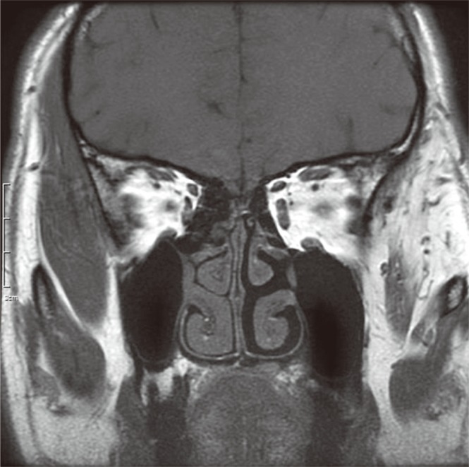
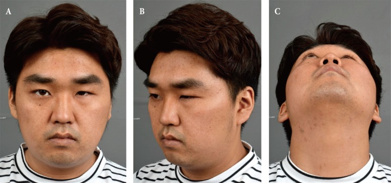
A 22-year-old man presented with a chief complaint of a one-year history of left temporal depression (Fig. 1). The patient had no history of craniofacial interventions or trauma. On physical examination, the patient had hollowing confined to the left temporal fossa. He did not exhibit facial expression and sensory abnormalities. Magnetic resonance imaging revealed fatty degeneration of the left temporalis muscle (Fig. 2). Other bony or soft tissue abnormalities were not reported. Electromyography of the facial nerve and nerve conduction studies on the trigeminal nerve performed to investigate the cause of fatty degeneration did not reveal any abnormalities. An autologous fat injection was planned to correct the hollowing deformity. The contour lines were marked to define the concavity of the left temple (Fig. 3). Autologous fat was harvested from the left thigh using the modified Coleman technique. Through two slit incisions on hair-bearing scalp, the harvested fat was injected into the subcutaneous fat layer using 1 mL syringes. Multiple injections and vigorous massage were performed to avoid bump or nodule formation. To compensate for the expected absorption, a total of 30 mL fat was injected as overcorrection and temporal hollowing was well corrected during the four-month follow-up (Figs. 4, 5).

DISCUSSION

Temporal hollowing is defined as a depression in the temporal region, an area confined superior to the zygomatic arch and immediately posterior to the lateral orbital rim [2]. Atrophy of the superficial temporal fat pad—caused by ischemia, denervation, or displacement through dissection, disinsertion, or atrophy of the temporalis muscle caused by injury to the frontal branch of the facial nerve—may result in temporal hollowing. Disruption of the suspensory ligament of the superficial temporal fat pad, the middle and deep temporal arteries, or the zygomaticotemporal nerve can also cause temporal fat pad atrophy and temporal hollowing [2]. Coronal approach surgery is the most common cause of temporal hollowing, with a reported incidence of up to 6% [3]. Lipopenia, which results from certain medications or diseases, and zygomaticotemporal fracture can also lead to temporal hollowing. The incidence of traumatic temporal hollowing without coronal approach surgery was reported to be 6.1% [4].
Augmentation using porous hydroxyapatite or titanium mesh can be a treatment option for temporal hollowing [4,5]. It has been reported that an aesthetically improved outcome can be obtained with artificial materials, without major complications such as recurrence of hollowing, infection, and protrusion through skin. However, some patients complain about the excessive rigidity of the temporal area. Autologous fat grafts can be an option for mild to moderate temporal hollowing, which obviates the need for long incisions, longer operative times, and postoperative palpable rigidity. Although it should be repeated and overcorrected because of fat absorption, it can be done under local or general anesthesia.
In the current case, the patient developed temporal hollowing but did not have a history of trauma, surgery, or myogenic disease. Little is known about the exact pathophysiology of cryptogenic temporal hollowing, which deserves further study.

Notes

No potential conflict of interest relevant to this article was reported.

References

1. Lacey M, Antonyshyn O, MacGregor JH. Temporal contour deformity after coronal flap elevation: an anatomical study. J Craniofac Surg 1994;5:223-227. PMID: 7833395.
crossref pmid
2. Baek RM, Heo CY, Lee SW. Temporal dissection technique that prevents temporal hollowing in coronal approach. J Craniofac Surg 2009;20:748-751. PMID: 19480035.
crossref pmid
3. Frodel JL, Marentette LJ. The coronal approach. Anatomic and technical considerations and morbidity. Arch Otolaryngol Head Neck Surg 1993 119:201. -207. discussion 140. PMID: 8427684.
crossref pmid
4. Guo J, Tian W, Long J, Gong H, Duan S, Tang W. A retrospective study of traumatic temporal hollowing and treatment with titanium mesh. Ann Plast Surg 2012;68:279-285. PMID: 21346531.
crossref pmid
5. Moreira-Gonzalez A, Jackson IT, Miyawaki T, DiNick V, Yavuzer R. Augmentation of the craniomaxillofacial region using porous hydroxyapatite granules. Plast Reconstr Surg 2003;111:1808-1817. PMID: 12711940.
crossref pmid
Fig. 1

(A) The frontal view of a 22-year-old man with left temporal depression. (B) The 45° lateral view of a 22-year-old man with left temporal depression. (C) The worm’s-eye view of a 22-year-old man with left temporal depression.

acfs-17-218-g001.jpg
Fig. 2

Preoperative magnetic resonance imaging shows fatty degeneration of left temporalis muscle.

acfs-17-218-g002.jpg
Fig. 3

(A) Intraoperative frontal view photo taken before autologous fat graft. The contour lines were marked to define the concavity of the left temple. (B) Intraoperative lateral view photo taken before autologous fat graft. The contour lines were marked to define the concavity of the left temple.

acfs-17-218-g003.jpg
Fig. 4

(A) Immediate postoperative frontal view photo taken after autologous fat injection to the left temple. (B) Immediate postoperative bird’s-eye view photo taken after autologous fat injection to the left temple. (C) Immediate postoperative lateral view photo taken after autologous fat injection to the left temple.

acfs-17-218-g004.jpg
Fig. 5

(A) Frontal view photograph taken four months after surgery. (B) 45° lateral view photograph taken four months after surgery. (C) Worm’s-eye view photograph taken four months after surgery.

acfs-17-218-g005.jpg
TOOLS
Share :
Facebook Twitter Linked In Google+ Line it
METRICS Graph View
  • 3 Crossref
  •   Scopus
  • 4,495 View
  • 72 Download
Related articles in ACFS


ABOUT
ARTICLE CATEGORY

Browse all articles >

BROWSE ARTICLES
AUTHOR INFORMATION
Editorial Office
Dept. of Plastic and Reconstructive Surgery Chonnam National University Medical School, 42 Jebong-ro, Dong-gu, Gwangju 61469, Korea
Tel: +82-62-220-6354    Fax: +82-62-220-6357    E-mail: office_acfs@kcpca.or.kr                

Copyright © 2024 by Korean Cleft Palate-Craniofacial Association.

Developed in M2PI

Close layer
prev next