Retained large glass fragments for over 40 years in the maxillofacial region
Article information
Abstract
Foreign body (FB) impaction in the maxillofacial area could be caused by knives, glass fragments, and vegetative materials. We present the rare case of a 62-year-old man with a large glass FB in the left cheek retained for over 40 years. He had traffic accident over 40 years ago and glass fragments impacted on his left cheek. Glass fragments were retained around the zygomatic arch with dimpled scar and unclear serous discharge, but other facial motor or sensory dysfunction was not observed. We confirmed three glass fragments with radiologic examination including plain radiograph and computed tomographic image. Under general anesthesia, impacted glass fragments were removed through the direct incision on the dimpled scar and the additional incision on the left lateral canthal area. Remnant FBs were not seen on an intraoperative C-arm radiograph. After 2 days of irrigation for inflammation control, the dimpled wound was sutured. The wound was healed without major complication and the original dimpled scar was much improved.
INTRODUCTION
Foreign body (FB) penetration can be caused by therapeutic interventions, vehicle accidents, gunshots, and other traumatic injuries in the head and neck region [1]. A wide range of impacted FBs have been reportedly found in the maxillofacial area including knives, glass fragments, and vegetative materials [2,3]. Glass-related wounds comprise 13% of traumatic wounds and it is the most commonly retained FB in the human body, accounting for 53% of FB-related legal claims [4]. We present the rare case of a 62-yearold man with a large glass FB in the left cheek retained for over 40 years. The wound was not accompanied by distinct complications such as cellulitis, gas gangrene, or facial nerve damage but had continuous serous discharge along with a dimpled scar and chronic inflammatory responses.
CASE REPORT
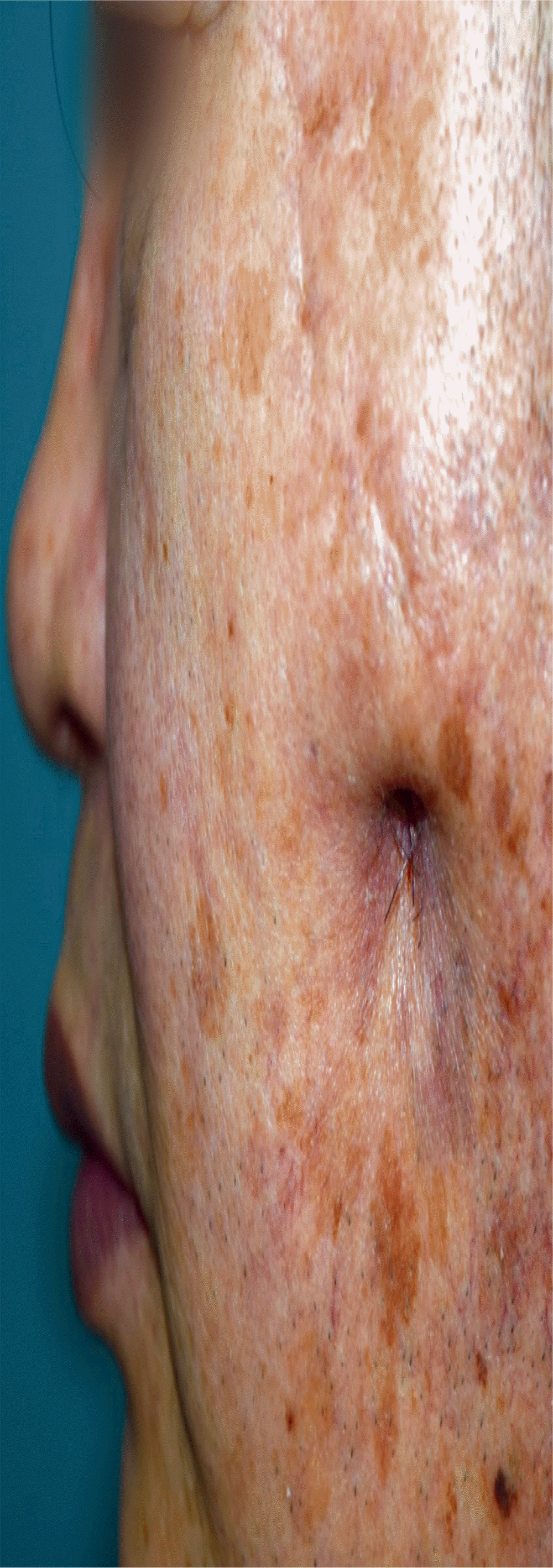
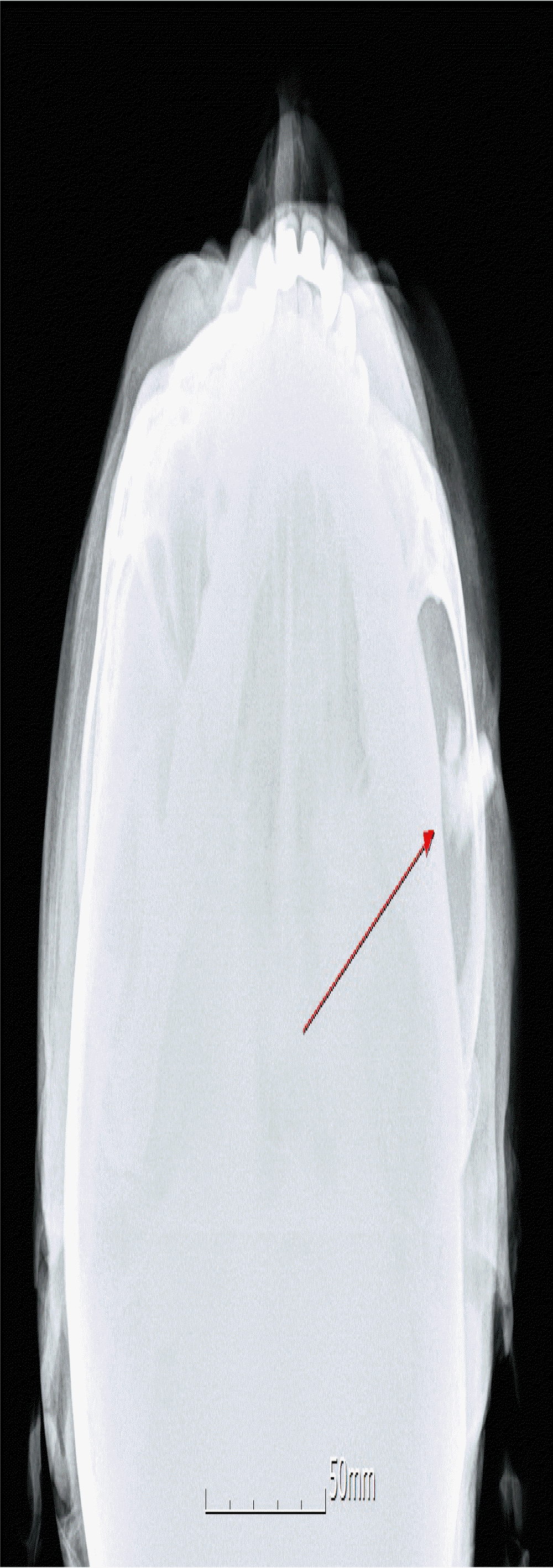
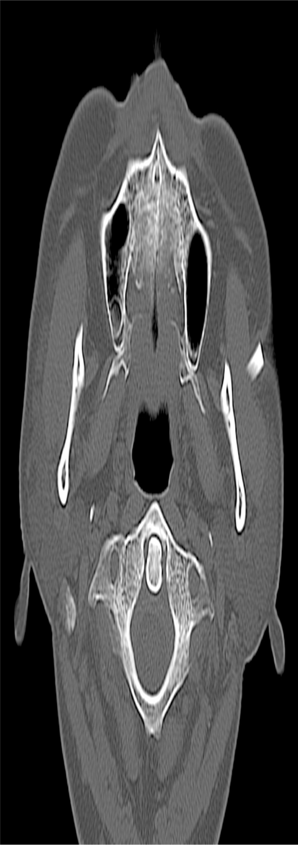
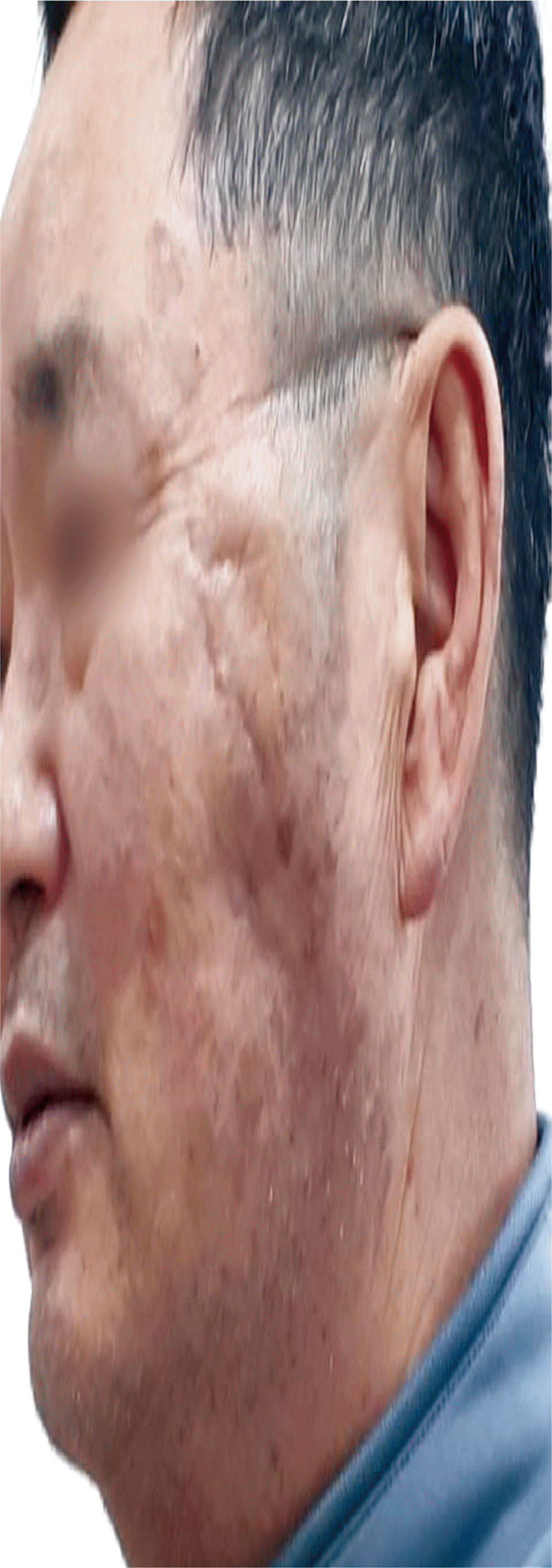
A 62-year-old man visited Hallym University Sacred Heart Hospital with a palpable mass on his left cheek. The patient had a history of traffic accident that had happened over 40 years before. Trials for the removal of the glass fragments were not executed because the patient did not manifest significant complications and only exhibited a dimpled scar on the left cheek and unclear serous discharge. Currently, a hard glass tip was palpated in the dimpled scar with unclear serous discharge on the left cheek (Fig. 1). Plain radiographic image showed three large pieces of glass (17.0 mm×6.5 mm, 5.5 mm×5.0 mm, 5.0 mm×5.0 mm, respectively) embedded around the left zygomatic arch (Fig. 2). Computed tomography (CT) confirmed the presence of three fragments of glass in the soft tissue. The largest and longest rectangular fragment was located just below the skin and palpated at the dimpled scar. The fragment penetrated superficial muscular aponeurotic system and impacted into the masseter muscle in front of the parotid gland, but the patient had intact cranial motor functions, such as facial animation, swallowing, phonation, and intact facial sensory function (Fig. 3). The other two fragments also were found below superficial muscular aponeurotic system and were located between the zygomatic arch and masseteric muscles. The dimpled skin shown on CT was slightly thickened, suggesting chronic inflammatory responses.
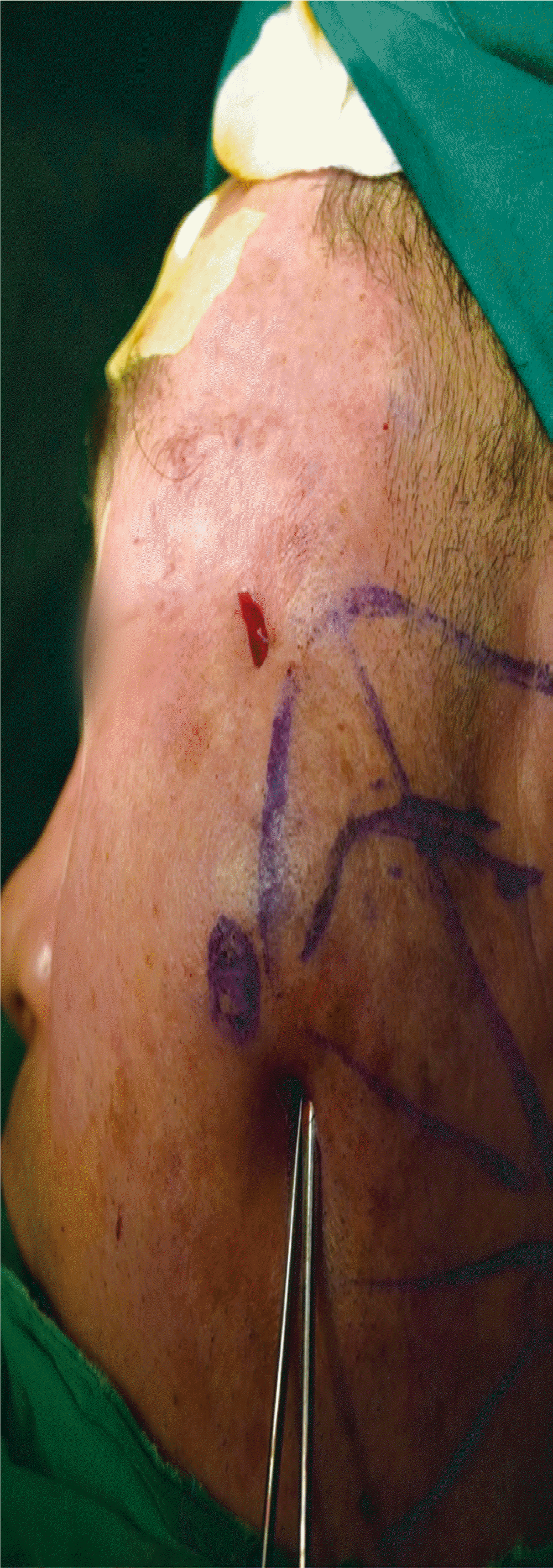
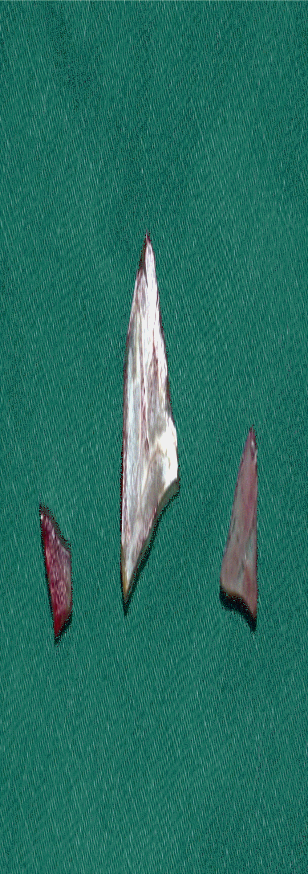
Exploration and impacted FB removal was performed under general anesthesia. Intraoperative C-arm radiography revealed three fragments of radio-opaque FBs. The dimpled wound was incised and the long rectangular glass fragment was removed. Fibrous capsule was formed from the masseter superficial surface to the incised skin which also exhibited cicatricial tissue with inflammatory responses. Branches of facial nerve were not found in the capsule. Superficial capsular tissue and inflammatory skin were debrided without total capsulectomy considering damage to zygomatic or buccal branch of facial nerve which would be located around the deep fascia. The other two fragments could not be approached through the scar incision without masseter muscle and deep fascia resection which could lead to facial nerve injury. In order to avoid iatrogenic facial nerve or parotid duct injury, a secondary incision with 1.5 cm length on the left lateral canthal area was made for exploration (Fig. 4). Exploration was performed through the plane beneath the masseter muscle and the two glass fragments were removed through the secondary incision (Fig. 5). Fibrous capsule was not found due to narrow view of secondary incision. Remnant FBs were not seen on an intraoperative C-arm radiograph. Copious saline irrigation was done through the incisions and the lateral canthal incision was first closed. The scar incision was opened for the tertiary closure after inflammation control.
Saline irrigation and the application of betadine-soaked dressing was performed on the cheek incision for 2 days and the inflammation, which was accompanied by unclear discharge, was alleviated. Under local anesthesia, scar tissue debridement and layer by layer closure were followed. The wound healed without complications. Postoperative facial motor dysfunction and sensory deficit were not observed. Postoperative 27-month photograph showed that the unsightly dimple on the left cheek was greatly improved, and only a slight remnant dimple remained, which could be attributed to remaining capsular contracture (Fig. 6).
DISCUSSION
FB impaction in the maxillofacial area is uncommon and a third of FBs are unnoticed in this region [5]. The types of FBs vary widely from nonvegetative materials, such as glass, metal bullets, and stones to vegetative materials [6]. Impacted FBs can cause complications, including infection with pain, swelling, peripheral nerve damage, pseudoaneurysm, and synovitis [6]. Foreign materials are often antigenic and produce chronic inflammatory responses [7]. The intensity of an inflammatory reaction is largely determined by the chemical composition and physical form of FBs [6]. The impacted glass in our case had a smooth, nonporous surface, and this lessened the inflammatory response in contrast to other vegetative FBs including wood, thorns, and spines [6]. However, prolonged inflammation associated with long-standing impacted FBs results in delayed wound healing and soft tissue and bone destruction [6,7]. Retained glass fragments in the head and neck region also reportedly cause a cutaneous fistula with unclear discharge, similar to our case [8].
Injuries of the head and neck region can damage neurovascular structures, including cranial nerves, the carotid arteries, and the jugular veins. This patient had a penetrating injury without signs and symptoms of cranial nerve dysfunction and vascular compromise but in the literature, vascular damage and signs of neurologic deficit, including swallowing difficulty, hoarseness, loss of tongue sensation, tongue deviation, and protrusion after FB penetration, have been reported [9,10]. Facial paralysis due to chronic inflammation associated with a retained FB has also been reported. The patient had three pieces of large glass in the left cheek accompanied by a dimpled scar with unclear discharge, which may be attributed to chronic inflammation [11]. The fragments were impacted in an extensive area around the left cheek. The fragments were positioned near vital structures, including the parotid gland and the masseter muscle. Due to the position of the glass particles, chronic inflammation exacerbation resulted from FB response to the large glass, which could damage adjacent structures, such as the parotid gland and duct, masticatory muscles, facial nerves, and superficial temporal artery if the glass remained intact for a longer period.
In conclusion, FBs in the maxillofacial region should be completely extracted because they cause deformities in the regional anatomy, subsequent scarring following inflammatory responses, and vital structure injuries. Furthermore, patients with cutaneous dimpled scar in this area should be evaluated by taking the history of previous trauma. Radiographic examinations including simple radiography and CT should also be performed.
Notes
No potential conflict of interest relevant to this article was reported.
Notes
PATIENT CONSENT
The patients provided written informed consent for the publication and the use of their images.
