Treatment of post-traumatic hematoma and fibrosis using hyaluronidase injection
Article information
Abstract
Patients complaining of swelling and hematoma caused by contusion of the face can be easily seen in the emergency room. Most of the treatments were conservative treatments such as ice bag application, mild compression dressing, and massage. During the follow-up, fibrosis progression due to hematoma was frequently observed in the contusion site. When hematoma or fibrosis is confirmed, hyaluronidase (H-lase) 1,500 IU and 2 mL of normal saline were mixed and subcutaneously injected in crisscross manner. To evaluate the improvement of hematoma before and after hyaluronidase injection, three plastic surgeons evaluated using the Vancouver scar scale and compared preoperative and postoperative images. Hematoma and fibrosis after facial trauma improved after hyaluronidase injection for early treatment.
INTRODUCTION
Swelling and hematoma due to facial contusion are one of the most common traumatic complications. Because swelling and hematoma are mostly absorbed by the observation, they are often not treated aggressively. However, if left untreated, hematoma can cause fibrosis. At that stage, cosmetic deterioration occurs, which can lead to psychological stress and social discomfort to the patients. Especially, hematoma develops often after facial contusion in patients taking anticoagulants, such as aspirin, clopidogrel, and warfarin for the prevention or treatment of heart or cerebrovascular disease, and unfortunately, conservative methods have their limitations. Hyaluronidase has the ability to break down the hyaluronic acid, facilitating the absorption of hematomas and edema through peripheral lymphatic channels and capillaries while the local intercellular permeability increase [1,2]. We report one case of hematoma and another case of fibrosis in which the patient’s clinical condition improved after administration of a hyaluronidase injection for the early treatment of hematoma or fibrosis after facial trauma.
CASE REPORTS
Case 1
A 66-year-old female patient visited the emergency room complaining of pain and swelling at the left periorbital area caused by contusion. There was no fracture. After applying ice bags for 2 days, the patient came to our outpatient department because the swelling was still present in the left periorbital area (Fig. 1). On physical examination, mild hematoma was observed. Hyaluronidase (H-lase; Kuhnil, Seoul, Korea) 1,500 IU and 2 mL normal saline were mixed, and it was injected subcutaneously with 30-gauge needle; then a compression massage was performed for 5 minutes.
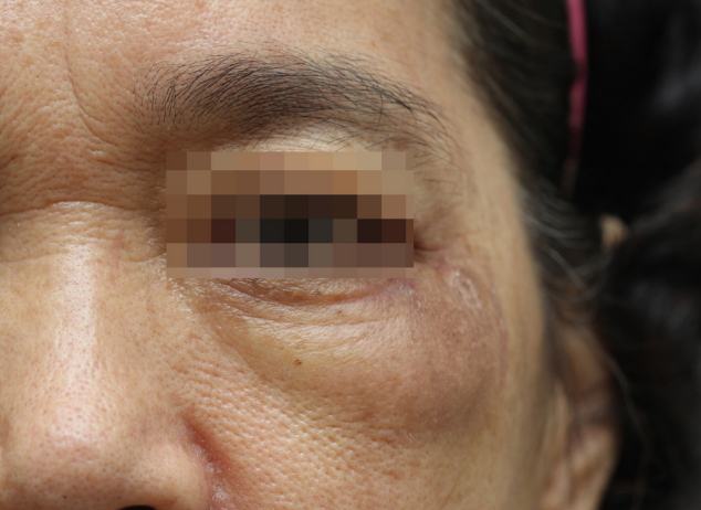
Frontal view of a 66-year-old woman with hematoma on the left periorbital area.
To determine the changes before and after hyaluronidase injection, three plastic surgeons (J Kim, KC Yoon, HW Shin) evaluated the patient, using the Vancouver scar scale (VSS), and compared preoperative and postoperative images. All photographs were taken by the same photographer, using Canon 700 D camera (Canon Inc., Tokyo, Japan) and the images were obtained under the same conditions to maintain consistency between the images. Photographs were taken at the same position, in the same posture, from the same angle with the same lighting conditions.
Hyaluronidase was injected three times at weekly intervals, and the effect was evaluated at 3 months after the last injection (Fig. 2). All hematomas were resolved, and there was no cosmetic deterioration at the contusion site. The VSS score improved significantly from 9 points to 1 point, and the patient was very satisfied with the treatment.
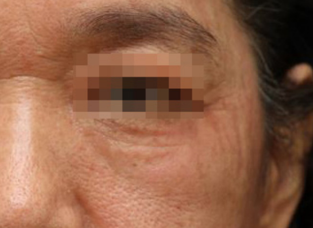
Frontal view photograph taken 3 months after three times’ hyaluronidase injections.
Case 2
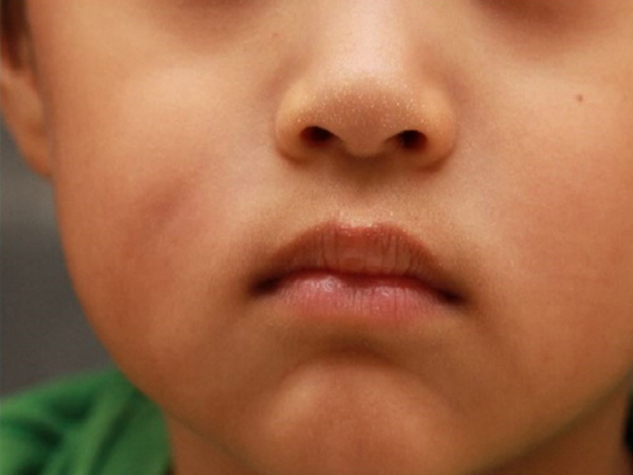
A 6-year-old male patient visited the emergency room complaining of pain on the right cheek area, which developed after he hit the corner of a piece of furniture. There was no evidence of fracture. After ice bag applications, the contusion site was observed at weekly intervals. One week after contusion, we observed dimpling in the right cheek area due to fibrosis (Figs. 3, 4).
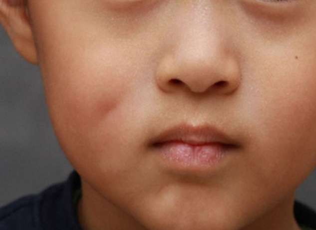
Frontal view of a 6-year-old boy with fibrosis on the right cheek area.
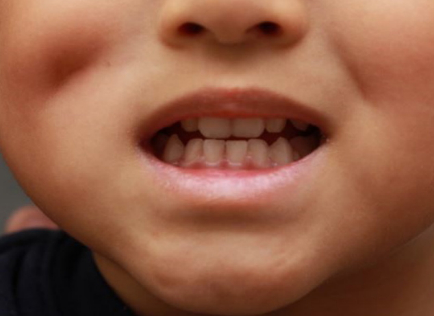
Frontal view of a 6-year-old boy with smiling.
Hyaluronidase 1,500 IU and 2 mL of normal saline were mixed and injected subcutaneously in a crisscross pattern. Compression massage was performed for 5 minutes after hyaluronidase injection. In total, four hyaluronidase injections were administered at weekly intervals.
The fibrosis, which was firmly felt by touching the right cheek area, softened and the dimpling improved noticeably. Three plastic surgeons evaluated the patient, using the VSS, and the VSS score improved from 5 points to 2 points. The patient and his parents were very satisfied with the treatment (Figs. 5, 6).
Frontal view photograph taken 3 months after four times’ hyaluronidase injections.

Frontal view photograph with smiling taken 3 months after four times’ hyaluronidase injections.
DISCUSSION
Hematoma is a localized collection of coagulated and solidified blood from damaged vessels, caused by disease or trauma; it may lead to complications, such as poor wound healing, localized infection, and skin necrosis. Therefore, a more aggressive treatment is needed when hematoma is confirmed. Various methods have been tried to treat hematoma and fibrosis caused by trauma; however, no effective method have been identified [1]. Herewith, we report observing significant improvements in a patient with hematoma and another patient with fibrosis by hyaluronidase injection. Swelling, hematoma, pain, and the sensation of itching were improved in these patients who received hyaluronidase.
At the time of treatment, hyaluronidase injection was able to rapidly liquefy the solidified hematoma, causing rapid absorption and loss of hematoma. The treatment period was much shorter than that of massage or observation (in case 1, hematoma disappeared after 2 weeks of hyaluronidase injection). This has also prevented fibrosis from long-term wound hematoma.
Hyaluronidase has been shown to break down hyaluronic acid, which is the ground substance of the connective tissue, by separating 1,4-glucosaminidic bond between C1 and the glucosamine moiety and C4 of the glucuronic acid [3]. Treatment with hyaluronidase is effective owing to increases in the diffusion and absorption rate, which in turn, increases the intercellular permeability [1,4,5]. No complications were observed after treatment with hyaluronidase. Fibrosis after trauma occurs because of the activation of fibroblasts, resulting in increased collagen synthesis and accumulation of thin and disorganized collagen fibers [5,6]. It is generally known that hyaluronidase inhibits fibroblast proliferation [7].
We injected hyaluronidase directly into the fibrosis site, and it degraded hyaluronic acid to lower the concentration of the intercellular ground substance [1], hence improving the status of pain, pliability and stiffness on the fibrosis site. The improvement of fibrosis may be due to physical dissection by the needle itself during injecting hyaluronidase by the crisscross method which results in an increase in the spacing between the fibrotic tissues, and hence, the improvement of fibrosis. The effect of hydro dissection as well as the hyaluronidase itself may have influenced the improvement of fibrosis.
However, adhesiolysis using a needle without the hyaluronidase injection stresses the internal scar and results in repeat adhesions [5]. To minimize needle-induced adhesiolysis, we used the thinnest needle (30-gauge needle). Injection of hyaluronidase into the fibrotic scar lesion was effective in improving the pliability, height, and pigmentation of the fibrotic lesions by breaking down the ground substance of the connective tissue [1].
The VSS is used primarily as an index of scar after external wound. In the absence of appropriate indicators for the judgment of hematoma or swelling, we compared the efficacy of hematoma and swelling using the VSS in this study. The VSS is evaluated on four scales: pigmentation, vascularity, pliability, and height. In hematoma, pigmentation, pliability, and height can be applied. In fibrosis, all four scales are improved in the treatment response, so we decided that it is appropriate to judge the treatment response. In mild hematoma and fibrosis cases, hyaluronidase injection may be considered as an alternative to hematoma removal treatment by surgical methods.
Notes
No potential conflict of interest relevant to this article was reported.
PATIENT CONSENT
The patients provided written informed consent for the publication and the use of their images.