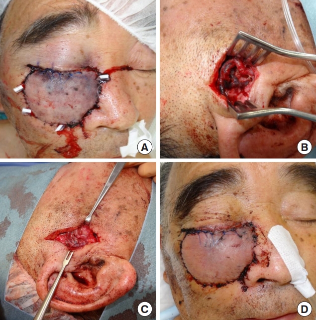
Small-diameter variation or angiectopia of the superficial temporal vein (STV) has been reported; however, anomalies at its entrance into the parotid gland have not been reported. An 80-year-old man underwent facial reconstruction using a radial forearm free-flap after extensive resection of his right lower eyelid and cheek due to basal cell carcinoma. The procedureŌĆÖs recipient vessels were the superficial temporal artery and two STV (diameter: 2.7 mm and 1.5 mm). Vascular anastomosis successfully performed. The following morning, the transferred flap showed severe congestion (
Fig. A). Venous thrombosis occurred 4 cm from the proximal of the anastomotic site (
Fig. B). Vascular reanastomosis alone was unable to resolve the severe venous thrombosis, and the STV outside the parotid gland appeared dilated with severe stenosis at its entrance. However, dissection with the release of the stenosis and further vascular reanastomosis successfully restored venous patency (
Fig. C). Fibrous bands causing external compression of the STV were divided. Flap congestion improved immediately and the flap survived completely (
Fig. D). The STV entrance into the parotid gland should be examined in both cases of proximal venous thrombosis with satisfactory anastomosis and unexplained dilatation of the dissected STV; preoperative ultrasound may be useful for evaluating the diameter of STV in the latter.


