Treatment of multiple craniofacial osteomas by endoscopic approach
Article information

Abstract
Osteomas are benign osteogenic neoplasms that usually occur as solitary craniofacial lesions. Multiple osteomas are rare, particularly those that do not occur as part of an associated syndrome. We report a case of a 72-year-old woman who presented with multiple bony protrusions over the forehead and scalp. She denied any diagnosis of syndromes that are known to be associated with osteomas. Surgical excision was performed by endoscopically resecting more than 30 osteomas of the frontal and bilateral parietal bones. Compared with conventional surgical excision through a direct incision, endoscopic-assisted surgery is a simple and effective method for the treatment of multiple craniofacial osteomas and is associated with excellent cosmetic outcomes and no neurovascular complications.
INTRODUCTION
Osteomas are benign osteogenic neoplasms, which occur secondary to the proliferation of compact or cancellous bone and are observed in 0.01% to 0.43% of the general population. Based on the origin of the tumor, osteomas are classified into central, peripheral, and extra-skeletal types [1-3]. Although most peripheral osteomas show benign characteristics, osteomas that increase in size, particularly those at prominent sites such as the face, necessitate surgical intervention. Osteomas usually present as solitary lesions as few reports in the available literature have described multiple osteomas [4]. We report a rare case of non-syndromic multiple peripheral osteomas of the craniofacial bones in a patient who was surgically treated with endoscopic-assisted excision.
CASE REPORT
A 72-year-old woman presented with a greater than 10-year history of multiple bony prominences of the forehead. Each mass measured approximately 1 cm in diameter, and palpation showed hard, immobile, non-tender, oval-shaped elevated lesions (Fig. 1A). The patient denied a history of trauma to the area, and no other cutaneous lesions were observed elsewhere on her body. She denied diagnosis of any syndromes or diseases, except for well-controlled chronic hypertension and diabetes. The endogastroduodenoscopy and colonoscopy performed to rule out Gardner’s syndrome did not reveal any abnormalities of the gastrointestinal system. Intraoral evaluation also did not reveal any dental abnormalities, such as impacted or unerupted teeth. The preoperative computed tomography of craniofacial bones revealed multiple, small well-circumscribed lesions with density matching that of the surrounding osseous tissue, suggesting multiple peripheral osteomas of more than 30 lesions (Fig. 2A and B), primarily involving the frontal bone, as well as the upper aspects of the bilateral parietal bones.
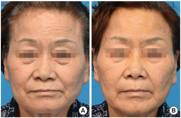
(A) Preoperative photograph of a 72-year-old female with multiple osteomas in the forehead. (B) Postoperative photograph at 10 days after the surgical treatment.

(A, B) More than 30 lesions of osteoma in the frontal and bilateral parietal bones are shown in the preoperative computed tomography (CT) scan. (C, D) The multiple osteomas were all removed except for the ones behind the hairline in the postoperative CT scan.
Using the monitored anesthesia care protocol, we made two small longitudinal incisions posterior to the hairline, and subperiosteal dissection was performed throughout the forehead via an endoscopic approach. Using osteotomes of varying degrees of curvature, we easily removed the well-defined masses, which were attached to the cortical bone, without destruction of the surrounding bone (Supplementary Video S1). The uneven surface was smoothened with a rasp to achieve a favorable contour of the forehead. The lesions posterior to the hairline that were cosmetically acceptable were left unremoved. A negative pressure drain was inserted through one of the incisions, and the wound was closed in layers.
The drain was removed on postoperative day 5, and the sutures were removed on postoperative day 10. We were able to observe aesthetically satisfactory outcomes without any postoperative complications, including hematoma, nerve injury, or any remnant palpable lumps. Histopathological examination confirmed mature lamellated bone fragments, consistent with osteomas. Photographs and computed tomography scans obtained 10 days postoperatively revealed a relatively well-smoothened surface of the forehead (Figs. 1B, 2C and D).
DISCUSSION
An osteoma is a relatively common type of osteogenic neoplasm of the craniofacial area, observed in 0.01% to 0.43% of the general population. These lesions develop secondary to the proliferation of compact or cancellous bone and are associated with multiple etiopathogenetic factors, including trauma, inflammation, developmental disorders, and genetic defects [3].
Osteomas usually occur as solitary lesions, and few cases of multiple osteomas have been reported in the previous literature [4]. Castelino et al. [5] described non-syndromic multiple gigantic osteomas involving the fronto-ethmoidal, mastoid, occipital, mandibular, and maxillary areas in a 21-year-old woman, who was successfully treated with surgical resection. Shin et al. [6] also reported a case of non-syndromic multiple osteomas in a 54-year-old woman who presented with more than 160 osteomas involving the frontal, parietal, and occipital bones. Notably, in most patients, multiple lesions occur as a clinical manifestation of Gardner’s syndrome, an inherited autosomal dominant condition that presents with colorectal polyposis, multiple osteomas, impacted or unerupted teeth, and cutaneous tumors. Bony lesions in patients with Gardner’s syndrome usually include mandibular osteomas; Utsunomiya and Nakamura [7] reported that among 15 families with familial polyposis coli, 27 of 29 patients presented with radiopaque mandibular lesions.
Our patient underwent endoscopic evaluation of the gastrointestinal tract to rule out Gardner’s syndrome; however, we observed no evidence of multiple polyposis. Gastrointestinal evaluation is essential for the assessment of multiple osteomas because non-syndromic multiple osteomas without underlying syndromes are rare and usually precede the presentation of colonic polyposis in Gardner’s syndrome. Therefore, craniofacial lesions can be considered sensitive markers for the early diagnosis of this condition [8].
Osteomas are classified into central, peripheral, and extraskeletal lesions, based on the origin and site of involvement [1,2]. Although most peripheral lesions present without accompanying symptoms, osteomas that occur over prominent areas, such as the forehead, often necessitate surgical removal owing to aesthetically unpleasant disfigurement of the face. Conventionally, forehead osteoma has been removed through direct incisions or via the coronal approach; however, endoscopic-assisted surgery has gained widespread acceptance for its efficacy and low complication rates [9-11].
No report in the available literature describes endoscopic-assisted resection of multiple craniofacial osteomas. The patient described in this study did not experience any symptoms associated with the osteomas, and surgery was performed solely for cosmetic correction of the facial disfigurement. For the best aesthetic results, two longitudinal minimal incisions were made posterior to the hairline to prevent visible postoperative scars. Moreover, the risk of accidental injury to the neurovascular structures of the forehead was minimized by making the incisions in longitudinal direction instead of transverse direction and by performing safe subperiosteal dissection under direct visualization of the field by the guidance of endoscopic light source. We observed no lumps of the forehead, hematoma, or nerve injury postoperatively, and the patient was satisfied with the results.
In conclusion, we report a rare case of non-syndromic multiple peripheral osteomas involving the craniofacial bones. Endoscopic-assisted surgery is a simple and effective approach for multiple craniofacial osteomas, with excellent cosmetic outcomes and minimal neurovascular complications.
Notes
Conflict of interest
No potential conflict of interest relevant to this article was reported.
Ethical approval
The study was approved by the Institutional Review Board of Seoul National University Bundang Hospital (IRB No. B-2008/631-101) and performed in accordance with the principles of the Declaration of Helsinki. Written informed consent was obtained.
Patient consent
The patient provided written informed consent for the publication and the use of her images.
Supplementary material
Supplemental Video S1.
The well-defined bony masses were easily removed without destructing the surrounding bone by using various degrees of curved osteotomes with the aid of visualization by endoscopic approach through the two minimal incisions behind the hairline.
Supplemental data can be found at: https://doi.org/10.7181/acfs.2020.00402.voo1.