Arch Craniofac Surg Search

CLOSE


Arch Craniofac Surg > Volume 23(2); 2022 > Article
Kim, Oh, Lee, and Yoon: Clinical association between serum cholesterol level and the size of xanthelasma palpebrarum

Abstract

Background

Xanthelasma palpebrarum (XP) is a benign periorbital lesion. The relationship between xanthelasma lesion size and serum cholesterol levels has been poorly studied. In this study, we investigated this relationship in the context of the clinical etiology of XP.

Methods

We retrospectively reviewed medical records and pathology reports of all patients treated for XP at our hospital between June 2014 and June 2021; the data were used to analyze lesion size, underlying disease, serum cholesterol, and disease recurrence.

Results

The mean values for patient age, serum cholesterol, and lesion size were 53.0 years, 235.0 mg/dL, and 69.2 mm2, respectively. XP mainly occurred in women (64.7%); furthermore, the incidence of XP and lesion size was greatest among patients in their 5th decade of life (41.2%). There was no statistically significant relationship between xanthelasma lesion size and serum cholesterol level.

Conclusion

This study compared lesion size with various clinical features in XP patients. In patients who underwent surgery for XP, serum cholesterol levels tended to be higher than those in the general population. However, the trend between the size of XP and serum cholesterol level was unclear. Therefore, if a patient with XP visits the hospital for surgery, it is recommended to check the lipid profile to confirm underlying dyslipidemia regardless of the size.

Abbreviations:

LDL,

low-density lipoprotein
;

XP,

xanthelasma palpebrarum
.

INTRODUCTION

Xanthelasma palpebrarum (XP) is a benign periorbital tumor. The term xanthelasma was first proposed by the 19th-century English surgeon and dermatologist, Sir Erasmus Wilson, derived from the Greek words “Xanthos” (yellow) and “Elasma” (beaten metal plate) [1]. As the name suggests, XP presents as yellowish papules or plaques on the eyelids.
The prevalence of XP is approximately 1.1% in women and 0.3% in men [2]. XP is known to be associated with hypercholesterolemia, and reports indicate that approximately 50% of patients diagnosed with XP also have hypercholesterolemia [2]. Histologically, xanthelasmas are composed of foam cells, which are lipid-laden histiocytes [3]. These foam cells accumulated mainly in the upper reticular dermis layer near the capillaries, appearing as yellowish skin lesions. Considering the pathogenesis, leakage of low-density lipoprotein (LDL) from the capillaries is an important aspect of the disease etiology [3,4]; mechanical movement and friction, heat, and inflammatory responses accelerate the leakage of LDL. These factors are prevalent around the eyelids, explaining the higher incidence of xanthelasma appearance compared other parts of the body [5].
Many studies have studied the clinical characteristics of xanthelasma, however, few studies have investigated the size of XP and the relationship between other clinical features. Therefore, in this study, we analyzed the size of XP lesions and their clinical characteristics to examine the relationship between elevated serum cholesterol levels and XP size.

METHODS

Study design and patients

We conducted a retrospective chart review of patients diagnosed with XP, who underwent surgery between August 2014 and September 2021. The participants of this study were patients with suspected XP lesions, confirmed by biopsy, in whom serum cholesterol levels were measured preoperatively. A total of 41 patients were clinically suspected of XP and underwent surgery. Four patients were excluded from the analysis because the pathologic diagnosis was not made. Three of the remaining 37 patients were excluded from the study because their serum cholesterol levels were not measured. A total of 34 patients were included in the analysis.

Evaluation of outcomes

We investigated the patient’s lesion size and location, serum total cholesterol level, underlying disease, and XP recurrence. Serum cholesterol levels were divided into normal (<200 mg/dL), intermediate (200–239 mg/dL), and hypercholesterolemia (≥240 mg/dL) groups. The area of the xanthelasma was assumed to have an elliptical shape and was calculated using the long (a) and short axis (b) measured preoperatively. If there were multiple lesions, the size of each lesion was added (Fig. 1).
Total lesions size of XP = iπ×ai2×bi2=iπaibi4
The lesions were classified according to the grading system proposed by Lee et al. in 2013 [6]. Lesions of grade I were limited to the upper eyelid, grade II involved the medial canthus, grade III affected both eyelids on the medial side, and grade IV involved both medial and lateral sides of the upper and lower eyelids (Fig. 2).

Operative technique

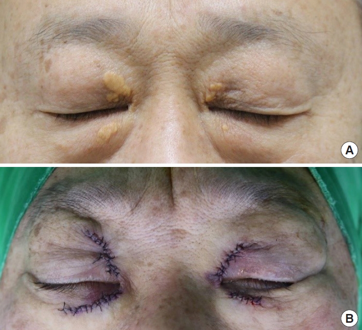
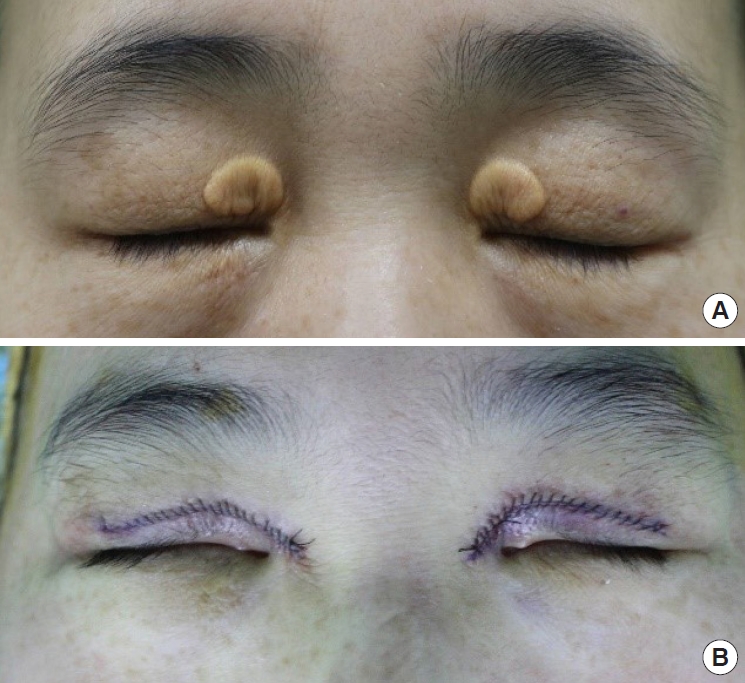
All patients underwent xanthelasma excision under local anesthesia. A simple excision of the lesion in an elliptical shape was generally performed to prevent eyelid deformities. In cases with multiple lesions, each lesion was excised through a different incision (Fig. 3). If the lesion had a diffuse distribution, a local flap or upper blepharoplasty was performed to prevent eyelid deformity (Fig. 4). Invasion of the xanthelasma into the orbicularis oculi prompted excision of the affected muscle section. The skin was repaired using black silk 7-0 sutures.

Statistical analysis

Continuous variables between groups were compared using Mann-Whitney U and Kruskal-Wallis tests. In addition, the one-sample Wilcoxon signed-rank test was used to compare the patients’ average serum cholesterol levels with those of the general population. Statistical significance was set at p<0.05. All analyses were performed using IBM SPSS Statistics for Windows (version 25.0; IBM Corp., Armonk, NY, USA).

RESULTS

Of the 34 patients analyzed, 12 were men (35.3%) and 22 were women (64.7%). The patients’ age at the time of surgery ranged from 34 to 74 years, with a mean age of 53.0 years. Grade I lesions were the most common (52.9%), followed by grades III (17.6%), II (14.7%), and IV (14.7%). Regarding the laterality of the lesion, 10 patients (29.4%) showed unilateral lesions and 24 patients (70.6%) showed bilateral lesions (Table 1).
The mean serum cholesterol level in all patients was 235.0 mg/dL. When the XP patients were classified by sex, serum cholesterol level was found to be 234.8 mg/dL in men and 235.2 mg/dL in women. As reported by a paper published in 2021 [7], the average cholesterol level in Korean adults was 192 mg/dL in men and 194 mg/dL in women. In this study, the serum cholesterol level was found to be higher in both men and women compared to the Korean general population (p=0.003 and p<0.001) (Fig. 5).
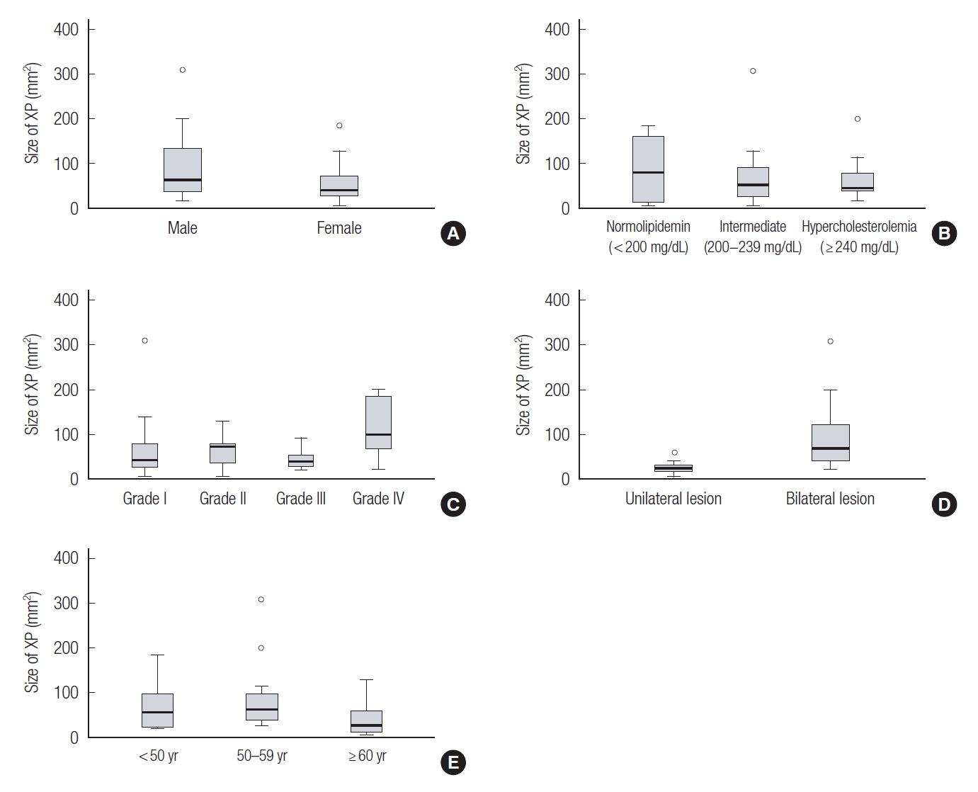
XP size was compared according to sex, grade, laterality, and serum cholesterol levels. The mean lesion was 69.2 mm2. According to the serum cholesterol level, the normal cholesterol group was largest size (87.0 mm2), followed by the intermediate group (69.8 mm2) and hypercholesterolemia group (63.1 mm2). However, the difference was not statistically significant. In addition, there were no significant differences in xanthelasma grade, age, or sex. The only significant difference was found in laterality, where bilateral lesions (87.8 mm2) were larger than unilateral lesions (24.7 mm2) (Table 2, Fig. 6).

DISCUSSION

This was a retrospective study investigating the relationship between XP size and other clinical characteristics. As in previous studies, XP was prevalent in middle-aged women [2,8]. In addition, grade I lesions involving only the upper eyelid were the most common, with the lesions coinciding on both sides. In this study, surgical excision was performed in all xanthelasma patients. Since the eyelids are the important structure that protects and lubricates the globes and cornea, it is essential to preserve their structure [9,10]. In most cases, simple excision followed by primary closure was enough. However, if the lesion had diffuse distribution, we performed local flap techniques or upper blepharoplasty in order to avoid eyelid deformity [4,6,11].
In papers published in Western countries, approximately 50% of patients with XP are associated with hypercholesterolemia [2]. Similarly, in a study conducted in Taiwan, dyslipidemia was present in 50% of patients [8]. This study found that approximately 38.2% of patients had hypercholesterolemia in the preoperative evaluation. This result is estimated to be higher than the prevalence of 20.7% in the general population of Korea [7].
Although there was no statistically significant difference in the size analysis of patients with XP, several trends could be identified. Most patients with XP ranged from 50 to 59 years of age, and the lesion size was the most extensive among these patients. XP is a more prevalent disease in women, but paradoxically, its size tends to be larger in men. It could be explained that women are more sensitive to facial aesthetics than men, so they are apt to come to the hospital for surgery.
A limitation to the generalizability of this study is that the sample size is relatively small and that it was conducted only at a single center. Therefore, a further large-scale study is needed. Another limitation was that other lipid profiles, including LDL cholesterol, were not examined. Meanwhile, in this study, comparing the serum cholesterol level with the general population requires careful interpretation. Since the analysis was performed on patients who underwent surgical treatment at the plastic surgery department, the possibility of selection bias should be considered. Also, in the case of patients taking lipidlowering medications, serum cholesterol may be lowered by the drug, so it is necessary to analyze them separately to account for this discrepancy. However, this study is limited in that it could not be implemented due to the omission of records, possibly leading to confounding bias.
This study compared lesion size with various clinical features in XP patients. In patients who underwent surgery for XP, blood cholesterol levels tended to be higher than those in the general population. However, the trend between the size of XP and serum cholesterol level was unclear. Therefore, if a patient with XP visits the hospital for surgery, it is recommended to check the lipid profile to confirm underlying dyslipidemia regardless of the size.

Notes

Conflict of interest

No potential conflict of interest relevant to this article was reported.

Ethical approval

The study was approved by the Institutional Review Board of Inje University Haeundae Paik Hospital (IRB No. HPIRB 2021-12-026-001) and performed in accordance with the principles of the Declaration of Helsinki. The requirement for written informed consent was waived.

Patient consent

The patients provided written informed consent for the publication and the use of their images.

Author contribution

Conceptualization: Young Geun Kim, Sung Ho Yoon. Data curation: Young Geun Kim. Methodology: Young Geun Kim, Sung Ho Yoon. Project administration: Sung Ho Yoon. Visualization: Young Geun Kim. Writing - original draft: Young Geun Kim. Writing - review & editing: Young Geun Kim, Jae Wook Oh, Keun Cheol Lee, Sung Ho Yoon. Investigation: Young Geun Kim. Supervision: Jae Wook Oh, Keun Cheol Lee, Sung Ho Yoon.

Fig. 1.
Xanthelasma palpebrarum-lesion size estimation methodology. The lengths of the major (a1 and a2) and minor (b1 and b2) axes were measured assuming the most similar ellipse, including that of the lesion. Based on this measurement, the sum of the ellipse areas was calculated to estimate the size of the lesion.
acfs-2022-00185f1.jpg
Fig. 2.
Xanthelasma grading system. (A) Grade I is limited to the upper eyelid; (B) grade II involves the medial canthus; (C) grade III affects both eyelids on the medial side; and (D) grade IV involves both the medial and lateral sides of the upper and lower eyelids.
acfs-2022-00185f2.jpg
Fig. 3.
A 59-year-old man with xanthelasma palpebrarum (grade IV). The patient was diagnosed with hypercholesterolemia and received lipid-lowering therapy. Lesions involving the upper and lower eyelids were observed on both sides and each was removed separately. (A) Preoperative and (B) postoperative clinical images.
acfs-2022-00185f3.jpg
Fig. 4.
A 49-year-old woman with xanthelasma palpebrarum (grade I). The patient reported that she had not been diagnosed with any disease. However, her total serum cholesterol level before surgery was 222 mg/dL, which led to her distribution in the intermediate cholesterol level group. Grade 1 lesions were present on both sides, and upper blepharoplasty was performed to prevent eyelid deformities. (A) Preoperative and (B) postoperative clinical images.
acfs-2022-00185f4.jpg
Fig. 5.
Comparison of serum cholesterol levels in xanthelasma palpebrarum (XP) patients and the general population. Regarding serum cholesterol levels, the mean serum cholesterol level in all patients was 235.0 mg/dL. When the XP patients were classified by sex, serum cholesterol level was found to be 234.8 mg/dL in men and 235.2 mg/dL in women, which was significantly higher than the average cholesterol concentration of Korean adults, 192 mg/dL in men and 194 mg/dL in women, respectively (one-sample Wilcoxon signed-rank test, p=0.003, p<0.001, respectively).
acfs-2022-00185f5.jpg
Fig. 6.
Comparison of serum cholesterol levels according to clinical characteristics. The size of the xanthelasma palpebrarum (XP) lesions of the patients was analyzed by dividing it into sex, total serum cholesterol, grade, laterality, and age. There were no statistically significant differences in sex (A), serum cholesterol concentration (B), grade (C), or age (E). Meanwhile, the bilateral lesion group had a significantly larger area when analyzed according to laterality (D).
acfs-2022-00185f6.jpg
Table 1.
Patient characteristics
Characteristics Value
Age (yr) 53.0 ± 8.2
 < 50 12 (35.3)
 50–59 14 (41.2)
 ≥ 60 8 (23.5)
Sex
 Male 12 (35.3)
 Female 22 (64.7)
Serum cholesterol level
 Normal (< 200 mg/dL) 4 (11.8)
 Intermediate (200–239 mg/dL) 17 (50.0)
 Hypercholesterolemia (≥ 240 mg/dL) 13 (38.2)
Xanthelasma grade
 Grade I 18 (52.9)
 Grade II 5 (14.7)
 Grade III 6 (17.6)
 Grade IV 5 (14.7)
Laterality
 Unilateral 10 (29.4)
 Bilateral 24 (70.6)
Past medical history
 Diabetes 1 (2.9)
 Hypertension 2 (5.9)
Recurrence 6 (17.6)
Body mass index (kg/m2) 23.9 ± 3.3
Size (mm2) 69.2 ± 64.4
Total serum cholesterol (mg/dL) 235.0 ± 38.6

Values are presented as mean±SD or number (%).

Table 2.
Size analysis of the xanthelasma palpebrarum
Variable Size of xanthelasma (mm2) p-value
Age (yr) 0.116a)
 < 50 67.2 ± 54.2
 50–59 87.6 ± 78.8
 ≥ 60 40.2 ± 41.7
Sex 0.168b)
 Male 95.8 ± 87.2
 Female 54.8 ± 43.7
Serum cholesterol level 0.971a)
 Normal (< 200 mg/dL) 87.0 ± 87.4
 Intermediate (200–239 mg/dL) 69.8 ± 72.0
 Hypercholesterolemia (≥ 240 mg/dL) 63.1 ± 49.2
Xanthelasma grade 0.431a)
 Grade I 66.8 ± 71.6
 Grade II 63.2 ± 46.7
 Grade III 44.2 ± 25.8
 Grade IV 114.0 ± 76.6
Laterality < 0.001b),c)
 Unilateral 24.7 ± 16.1
 Bilateral 87.8 ± 68.0

Values are presented as mean±SD.

a) Kruskal-Wallis test;

b) Mann-Whitney U test;

c) Statistical significance was set at p<0.05.

REFERENCES

1. Wilson E. Xanthelasma or xanthoma. Br Med J 1877;1:184.
crossref
2. Rohrich RJ, Janis JE, Pownell PH. Xanthelasma palpebrarum: a review and current management principles. Plast Reconstr Surg 2002;110:1310-4.
crossref pmid
3. Bergman R. The pathogenesis and clinical significance of xanthelasma palpebrarum. Acad Dermatol 1994;30(2 Pt 1):236-42.
crossref
4. Pe’er L, Nemet AY. Xanthelasma palpebrarum: an oculoplastic viewpoint of optimal treatment. Adv Ophthalmol Optom 2021;6:341-56.
crossref
5. Chung YH, Kang SY, Choi WS. A case of intramuscular xanthelasma palpebrarum found during blepharoplasty. Arch Craniofac Surg 2018;19:296-9.
crossref pmid pmc
6. Lee HY, Jin US, Minn KW, Park YO. Outcomes of surgical management of xanthelasma palpebrarum. Arch Plast Surg 2013;40:380-6.
crossref pmid pmc
7. Cho SMJ, Lee H, Lee HH, Baek J, Heo JE, Joo HJ, et al. Dyslipidemia fact sheets in Korea 2020: an analysis of nationwide population-based data. J Lipid Atheroscler 2021;10:202-9.
crossref pmid pmc
8. Wang KY, Hsu KC, Liu WC, Yang KC, Chen LW. Relationship between xanthelasma palpebrarum and hyperlipidemia. Ann Plast Surg 2018;80(2S Suppl 1):S84-6.
crossref pmid
9. Kim MJ, Oh TS. Treatment for ophthalmic paralysis: functional and aesthetic optimization. Arch Craniofac Surg 2019;20:3-9.
crossref pmid pmc
10. Lee JH, Woo SS, Shin SH, Kim HJ, Kim JH, Kim SH, et al. Upper eyelid reconstruction using a combination of a nasal septal chondromucosal graft and a Fricke flap: a case report. Arch Craniofac Surg 2021;22:204-8.
crossref pmid pmc
11. Chung CM, Tak SW, Lim H, Cho SH, Lee JW. Early cicatricial lagophthalmos release with pentagonal wedge resection of the scar, fat redistribution, and full-thickness skin grafting. Arch Craniofac Surg 2020;21:49-52.
crossref pmid pmc
TOOLS
Share :
Facebook Twitter Linked In Google+ Line it
METRICS Graph View
  • 2 Crossref
  •   Scopus
  • 4,005 View
  • 116 Download
Related articles in ACFS


ABOUT
ARTICLE CATEGORY

Browse all articles >

BROWSE ARTICLES
AUTHOR INFORMATION
Editorial Office
Dept. of Plastic and Reconstructive Surgery Chonnam National University Medical School, 42 Jebong-ro, Dong-gu, Gwangju 61469, Korea
Tel: +82-62-220-6354    Fax: +82-62-220-6357    E-mail: office_acfs@kcpca.or.kr                

Copyright © 2024 by Korean Cleft Palate-Craniofacial Association.

Developed in M2PI

Close layer
prev next