INTRODUCTION
Osteomas are slow-growing, benign, and osteogenic tumors. Its incidence rate has been reported to range from 0.014% to 0.43% [1,2]. Osteoma can be classified as central, peripheral, and extraskeletal. Central osteomas occur in the endosteum, peripheral osteomas in the periosteum, and extraskeletal osteomas in the muscle [3]. Cranial osteomas can be classified into intracranial intraparenchymal osteomas (the rarest form), skull base osteomas (the most common form), and cranial vault osteomas, according to their anatomical location [4]. Peripheral osteomas most commonly occur in the frontal, ethmoid, and maxillary sinuses [5]. In plastic surgery, peripheral osteomas arising from the frontal bone are mainly encountered clinically. Frontal peripheral osteoma presents as a single, solid, and immobile mass. In general, there are no other symptoms other than aesthetic problems, and if the size is large, symptoms such as headache and facial pain may appear [2,6]. This study aimed to analyze the clinical data of frontal peripheral osteomas through a retrospective study.
METHODS
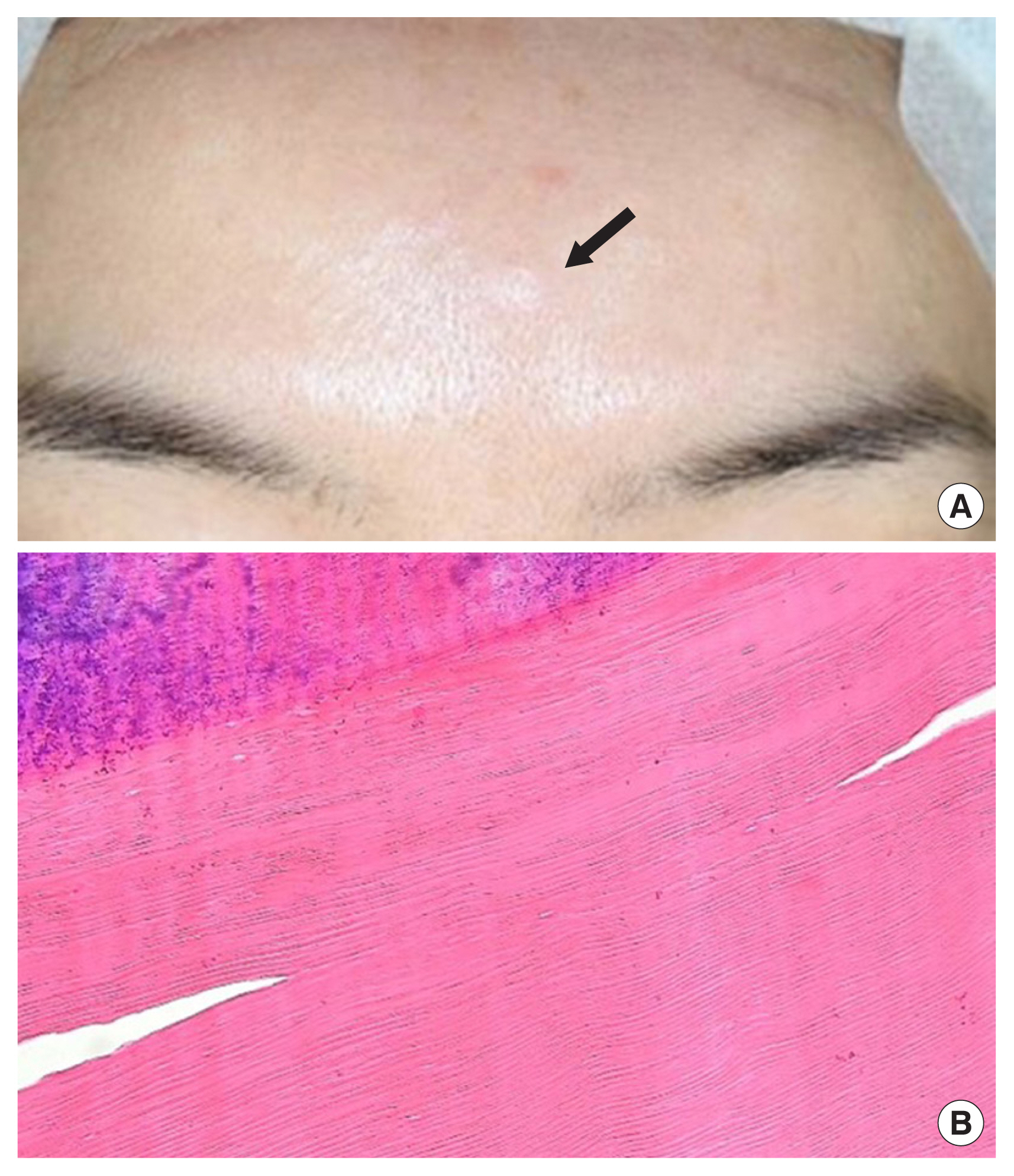
We retrospectively reviewed the medical records of patients who visited our hospital with frontal peripheral osteomas between January 2014 and June 2022. We analyzed the following variables: age, sex, type, single or multiple, size, history of head trauma, operation, and recurrence. It was classified into sessile and pedunculated types according to the shape detected on facial bone computed tomography (Fig. 1).
RESULTS
A total of 39 patients and 41 osteomas were analyzed. Their ages ranged from 4 to 78 years with a mean age of 52 years. A 4-year-old patient presented to our hospital after being diagnosed with osteoma at another hospital; however, her parents refused surgery. Among the patients, seven (18%) were men and 32 (82%) were women, with a man-to-woman ratio of 1:4.6. Two patients (5%) had multiple masses, with two osteomas in each, whereas only two patients (5%) had a history of head trauma. The first patient had a history of contusion at the same location 1 year prior to the onset of the osteoma. The second patient had a history of parietal bone fracture due to a fall 7 years before the onset of osteoma, and osteoma occurred in the frontal bone. A total of 29 patients (74%) underwent surgery, i.e., ostectomy, through a direct approach. None of the patients experienced recurrence. Follow-up was conducted for 6 months to 3 years. Twenty-nine osteomas (71%) were sessile, whereas 12 (29%) were pedunculated, and their size ranged from 4 to 30 mm, with an average of 10 mm (Table 1, Fig. 2).
DISCUSSION
The pathogenesis of peripheral osteomas remains controversial. Several theories have been proposed, including traumatic, infectious, and embryological causes [2,7ŌĆō9]. The traumatic theory relates to the fact that the sequestered nests may have been separated by a previous head trauma [2]. The embryologic theory states that osteomas arise from embryologic cartilaginous rests or embryologic periosteum [8]. Richards et al. [9] have suggested that osteomas can be stimulated by trauma, infection, or overgrowth of normal bony tubercles. Peripheral osteomas must be distinguished from exostoses of the jaws, which are bony excrescences that occur on the buccal side of the alveolar bone. These exostoses are believed to be of either reactive or developmental origin. Moreover, osteomas require differential diagnosis from osteoblastomas, osteoid osteomas, odontomas, and focal sclerosing osteomyelitis [10].
Patients with osteomas should be suspected of having Gardner syndrome. Colorectal polyposis, skeletal abnormalities, and multiple impacted or supernumerary teeth are triads of Gardner syndrome. Onset occurs in the second decade of life, with malignant transformation of colorectal polyps reaching 100% by the age of 40. Skeletal involvement includes both peripheral and endosteal osteomas, which can occur in any bone, but are found more frequently in the skull, ethmoid sinuses, mandible, and maxilla [11].
Osteomas appear as well-circumscribed, with well-defined radiopacity, on radiographic findings; computed tomography is the best modality for diagnosing osteomas [12,13]. Peripheral osteomas can be divided into two types based on their shape and pathological findings. Compact or ŌĆ£ivoryŌĆØ osteoma usually has a sessile base, normal-appearing dense bone with minimal marrow spaces, and occasional haversian canals. The size of different types of osteomas range from several millimeters to several centimeters; however, part of the lesion may be in the bone, masking the true size. Cancellous osteoma is usually pedunculated and resembles the bone of origin. It contains the trabeculae of the bone and fibrofatty marrow with osteoblasts. The surface can be irregular or smooth, with cortical bone at the margins [2].
The treatment of frontal peripheral osteomas involves surgical excision. There are direct open and endoscopic approaches to surgical excision [2,6,14,15]. Endoscopic removal of frontal osteomas was first reported in 1995. The endoscopic approach has the advantage of minimizing scarring and providing better aesthetic results. However, its disadvantages are a wider dissection than the direct approach, more swelling, longer postoperative recovery time, possibility of normal tissue damage during burring for removal, and time taken for the surgeon to learn the surgical technique [6,14].
We performed excision of frontal peripheral osteomas through direct approach in all patients, and burring was performed when irregularity was observed in the base margin after ostectomy. There were no particular complications or recurrences.
Our study analyzed a larger number of patients than other studies on frontal peripheral osteomas. We expect that the epidemiologic data of our study can help surgeons who encounter frontal peripheral osteomas in the field provide proper management for their patients.