Arch Craniofac Surg Search

CLOSE


Arch Craniofac Surg > Volume 24(2); 2023 > Article
Park, Lee, Kim, and Kim: Long-term outcomes after core extirpation of fibrous dysplasia of the zygomaticomaxillary region

Abstract

Background

Fibrous dysplasia (FD) is a localized bone disorder in which fibro-osseous tissue replaces the normal bone structure. Patients with craniofacial FD often present with gradual swelling, deformity, and compromised vision or hearing. We previously introduced “the core extirpation method,” a novel surgical technique that is minimally invasive like traditional bone shaving but has longer-lasting effects. This study presents the long-term outcomes of our core extirpation method.

Methods

We conducted a retrospective analysis of patients who underwent core extirpation for FD of the zygomaticomaxillary region from 2012 through 2021. Computed tomography (CT) scans were performed 6 to 12 months before the operation, immediately before and after the operation, and during follow-up visits. We performed all operations using the upper gingivobuccal approach, and we extirpated the core of the lesion while preserving the cortical structures of the zygoma and the maxilla to maintain symmetrical facial contour.

Results

In 12 patients with lesions in the growth phase, anteroposterior/mediolateral (AP/ML) length discrepancies and the volume increased between preoperative and immediate postoperative CT scans. All patients’ immediate postoperative AP/ML discrepancies were stable up to 12–17 months postoperatively. Postoperative volume showed continuous lesion growth; the median volume growth rate was 0.61 cc per month.

Conclusion

In this article, we present our experiences managing FD using the minimally invasive core extirpation technique, which entails small expected blood loss and can be performed as day surgery. It provides similar cosmetic outcomes as traditional bone shaving but with longer-lasting results. Although there are some limitations with the study’s retrospective nature and small sample size, our 4-year follow-up results show promising results of the core extirpation method in well-indicated patients.

Abbreviations:

AP/ML,

anteroposterior/mediolateral
;

CT,

computed tomography
;

FD,

fibrous dysplasia
.

INTRODUCTION

Fibrous dysplasia (FD) is a localized bone disorder that replaces normal bone structure with fibro-osseous tissue. FD accounts for approximately 6% of benign bone tumors, and patients affected in the craniofacial region often experience gradual swelling, deformity, and compromised vision or hearing [13]. While the natural disease course can vary between patients, there exist three phases of FD progression: initial, growth or proliferation, and stabilization [4,5].
Surgery was previously indicated for FD lesions in the active growth phase that caused neurological, functional, or aesthetic concerns due to its invasiveness [3]. The two main surgical treatments are radical excision with reconstruction and conservative bone shaving. While radical excision is less prone to regrowth, it is invasive, has high expected blood loss, and has a longer healing time [68]. On the other hand, conservative bone shaving provides relatively short-lasting results requiring reoperation every 2 to 4 years after surgery, and sometimes aesthetic concerns resurface after only a few months [9].
We previously introduced “the core extirpation method,” a novel surgical technique that is minimally invasive like traditional bone shaving but has longer-lasting effects [10]. Physical evaluations during clinical follow-up visits after the core extirpation technique in the zygomaticomaxillary region revealed no visible external regrowth during several years of monitoring. However, on computed tomography (CT) scans, refilling of the maxillary sinus by the remnant FD lesion was evident. This study presents the long-term outcomes of the core extirpation technique in managing zygomaticomaxillary FD.

METHODS

We retrospectively analyzed patients who underwent core extirpation for FD of the zygomaticomaxillary region between 2012 and 2021 at Seoul National University Bundang Hospital. This study was approved by the Ethics Committee of Seoul National University Bundang Hospital (IRB No. B-2104-676-103). Patient demographics, including age, sex, laterality, location, and initial symptoms, were compared. In this retrospective analysis, CT scans from each patient were analyzed chronologically.
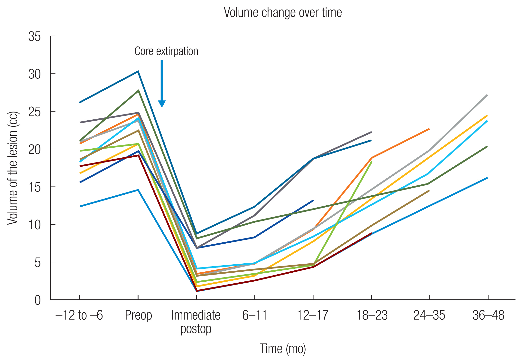
CT scans taken 6 to 12 months prior to the operation, immediately before and after the operation, and during follow-up visits were analyzed. Axial scans were used to measure the most prominent points of the zygomatic bone and to assess the discrepancies between the anteroposterior (AP) and mediolateral (ML) lengths between the affected and the unaffected sides (Fig. 1). Additionally, the volume of the FD lesion was manually measured from the axial cuts obtained in each scan.
All operations were performed under general anesthesia with the patient in the supine position and the head elevated. The upper gingivobuccal approach was utilized for subperiosteal dissection of the affected region to expose the lesion. A port in the anterior maxillary wall was created with a sagittal saw based on the preoperative CT scan, allowing access to the core of the lesion. The core of the lesion was extirpated while preserving the cortical structures of the zygoma and the maxilla to maintain symmetrical facial contour and improve aesthetic outcomes. Following core extirpation, the anterior maxillary wall and the zygomaticomaxillary complex were fixed using a miniplate if necessary to restore the buttresses. Further details of the core extirpation technique can be found in our previously published work [10].

RESULTS

A total of 12 patients (seven females and five males) were analyzed in this study. The mean age of the patients was 23.7 years (range, 11–40 years). Ten Patients presented with painless swelling of the facial area, and FD was incidentally diagnosed in two patients during a CT scan. Five patients were affected on the right side, while seven were on the left. Follow-up periods ranged from 12 to 48 months, with a mean follow-up time of 28.8 months (Table 1).
Chronological changes in the AP/ML discrepancies and the lesion volumes are summarized in Table 2 and Fig. 2. All 12 patients exhibited an increase in AP/ML discrepancies and volume from 6 to 12 months before the operation and immediate postoperative CT, indicating that all lesions were in the growth phase. The immediate postoperative AP/ML discrepancies were stable up to 12–17 months postoperative in all patients. One out of seven patients with postoperative 18–23 months CT scans (patient 3) had a slightly increased ML discrepancy from −1.1 to −0.5 mm, while the discrepancies were stable in the rest of the patients. In five patients with postoperative 24 to 35 months CT scans, the same patient (patient 3) showed increased ML discrepancy, while the other four had stable discrepancies. In four out of five patients with postoperative 36 to 48 months CT scans, AP/ML discrepancies increased. At 36 to 48 months, the median increase from immediate postoperative measurements was 0.78 mm in AP (range, 0–2.7 mm) and 2.26 mm in ML (range, 0.1–8.7 mm). Postoperative volumes showed continuous growth of the lesion, with a median volume growth rate was 0.61 cc per month (range, 0.33–0.89 cc). The total volume exceeded preoperative volume in three out of five patients with postoperative 36 to 48 months follow-up CT scans.
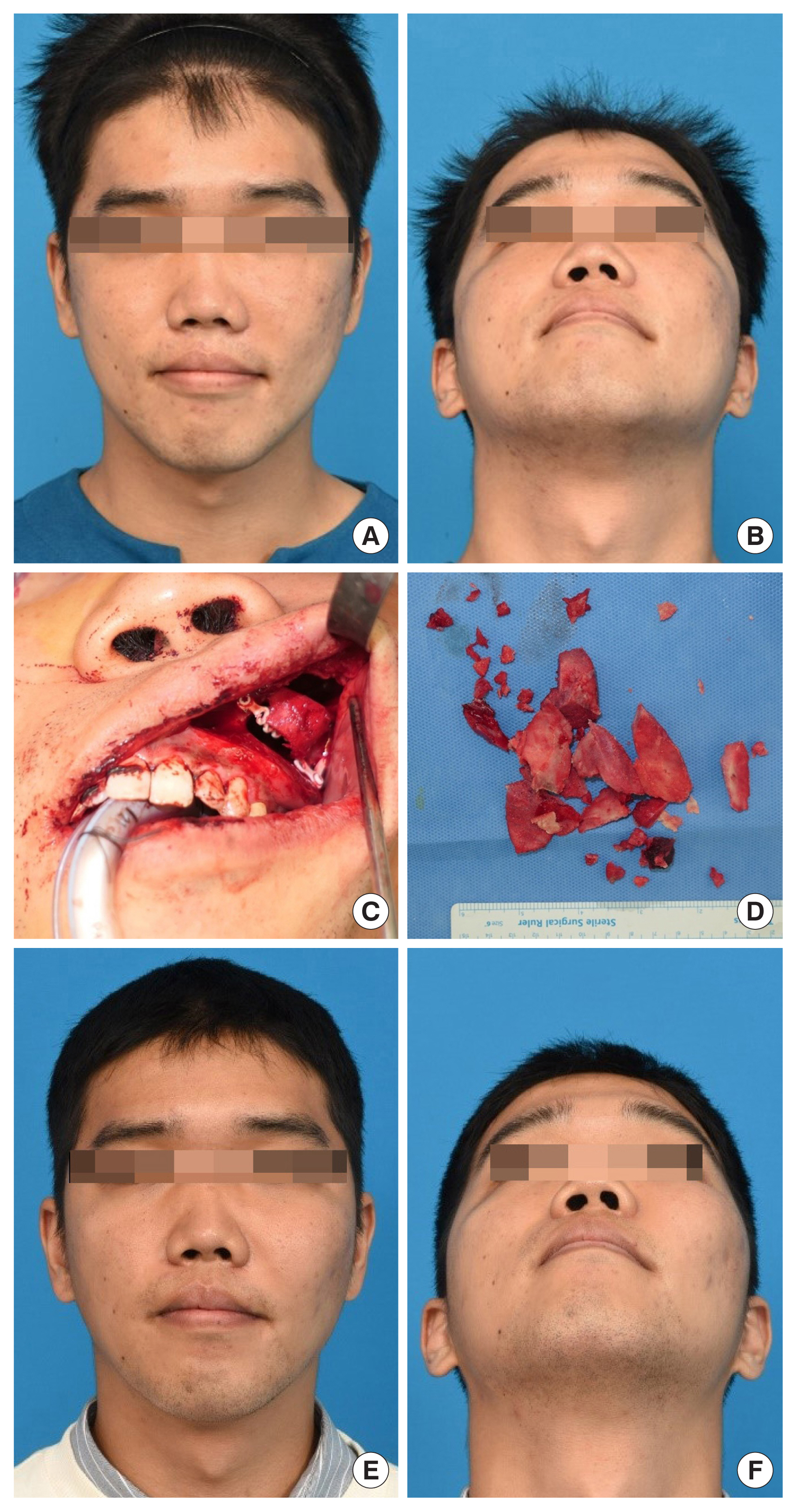
Representative case

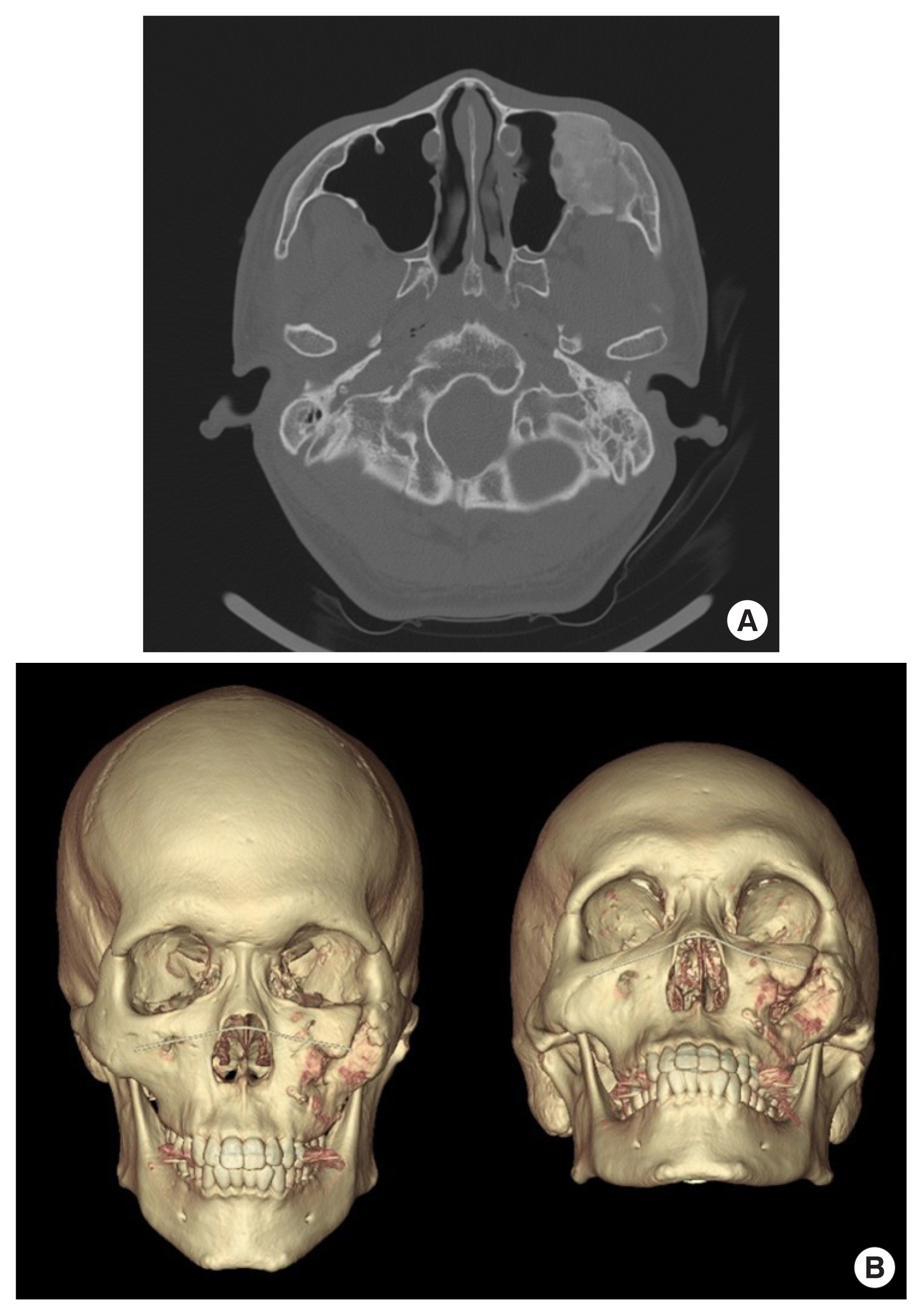
A 28-year-old man presented with a painless slowly-progressing swelling of the left cheek (Fig. 3). He had no signs of vertical dystopia, exophthalmos, or optic nerve compression. A CT scan demonstrated FD of the left zygomaticomaxillary complex, with preoperative AP and ML discrepancies of 8.39 mm and 6.5 mm, respectively. The patient underwent core extirpation with zygoma reduction without any postoperative complications. Although postoperative CT scans at 12 months showed intralesional growth, there was no further external increase in the lesion nor any evidence of recurrence (Fig. 4).

DISCUSSION

FD of the zygomaticomaxillary complex lesion can result in facial asymmetry due to bulging. Available surgical treatments for this condition include radical excision with immediate reconstruction and shaving [11]. Despite the relatively short effectiveness of bone shaving, many surgeons resort to bone shaving due to the difficulty and invasiveness of complete wide resection, which often requires complex reconstruction [12,13]. Several studies have reported excellent cosmetic outcomes with bone shaving [14,15], although the long-term efficacy of this technique remains controversial [16,17].
To achieve longer-lasting effects while minimizing invasiveness, we employed the core extirpation method. This minimally invasive technique is associated with low expected blood loss and can be performed as day surgery, providing similar cosmetic outcomes as traditional bone shaving but with longer-lasting results. Our results demonstrated that AP/ML dimensions were stable up to 24–35 months in all patients and that even after 36–48 months, the changes in AP/ML dimensions were minimal (average of 2.26 mm) and often went unnoticed by the patients.
The pattern of regrowth following the initial operation may account for the longer-lasting effect of the core extirpation compared to bone shaving. In the case of traditional bone shaving, the regrowth of the lesion occurs outward, resulting in external changes that can manifest within a few months. In contrast, following core extirpation, the remnant lesion regrows within the hollowed-out (extirpated) core area, with outward regrowth occurring only after complete filling of the core.
Radical excision followed by immediate reconstruction is a viable treatment option to eradicate FD lesions and prevent recurrences. This approach is recommended for patients with fast-growing lesions causing neurological or optical symptoms. However, in patients without these symptoms, the core extirpation method can be a suitable surgical option, especially in patients who do not want extensive surgery.
Although the core extirpation method cannot completely eradicate the lesion or halt its progression, our results showed that AP/ML discrepancies remained stable for up to 4 years after the initial operation. Therefore, routine CT scans for lesion follow-up may not be necessary during the first 4 to 5 years unless the patient reports symptoms or noticeable facial contour changes. This is particularly important in pediatric patients who are more sensitive to radiation exposure.
Several limitations should be acknowledged in this study. First, the sample size was small, which precluded statistical analysis. In addition, the retrospective design of the study is a further limitation. Future randomized studies with longer follow-up periods are necessary to fully evaluate the long-term effectiveness of the core extirpation method. In conclusion, our preliminary results after 4 years of follow-up indicate that the core extirpation method may be a promising alternative for well-selected patients.

Notes

Conflict of interest

No potential conflict of interest relevant to this article was reported.

Ethical approval

The study was approved by the Institutional Review Board of Seoul National University Bundang Hospital (IRB No. B-2104-676-103) and performed in accordance with the principles of the Declaration of Helsinki. The informed consent was waived because this study design is a retrospective chart review.

Patient consent

The patient provided written informed consent for the publication and use of their images.

Author contribution

Conceptualization: Joseph Kyu-hyung Park, Se Yeon Lee, Jong-Ho Kim, Baek-kyu Kim. Data curation: Joseph Kyu-hyung Park, Se Yeon Lee, Jong-Ho Kim. Formal analysis: Se Yeon Lee, Jong-Ho Kim. Methodology: Joseph Kyu-hyung Park. Project administration: Joseph Kyu-hyung Park. Visualization: Joseph Kyu-hyung Park. Writing - original draft: Joseph Kyu-hyung Park, Se Yeon Lee. Writing - review & editing: Joseph Kyu-hyung Park, Se Yeon Lee. Investigation: Joseph Kyu-hyung Park, Se Yeon Lee. Resources: Se Yeon Lee. Supervision: Joseph Kyu-hyung Park, Baek-kyu Kim. Validation: Baek-kyu Kim.

Funding

None.

Fig. 1
(A) Measurement of anteroposterior and mediolateral discrepancy (red line: anteroposterior discrepancy at the most prominent point of zygomatic bone/blue line: mediolateral discrepancy at the most prominent point of zygomatic bone). (B) Preoperative three-dimensional reconstruction image of computed tomography scan.
acfs-2023-00038f1.jpg
Fig. 2
Changes in the volume of the fibrous dysplasia lesion before and after surgery. The 12 patients included in the study showed an increase in anteroposterior/mediolateral (AP/ML) discrepancies and lesion volume on preoperative and immediate preoperative computed tomography (CT) scans. However, all patients exhibited stable AP/ML discrepancies up to 12–17 months postoperatively. Postoperative CT scans showed continuous growth of the remnant lesion, with a median volume growth rate of 0.61 cc/month.
acfs-2023-00038f2.jpg
Fig. 3
A 28-year-old man with painless swelling of left zygomatic area. (A, B) Preoperative photograph. Intraoperative photograph showing core extirpation using intraoral approach (C) and macroscopic specimen showing extirpated lesion (D). (E, F) Postoperative photograph showing no evidence of external increase of the lesion or recurrence.
acfs-2023-00038f3.jpg
Fig. 4
Postoperative 12-month computed tomography imaging of a representative case. Core extirpation with zygoma reduction was performed without any postoperative complications. (A) Axial cut showed intralesional growth, but (B) three-dimensional reconstruction image indicates no evidence of recurrence or external increase of the lesion beyond the preoperative contour.
acfs-2023-00038f4.jpg
Table 1
Patient demographics
Case Age (yr) Sex Laterality Location Initial symptom Follow-up (mo)
1 19 F Left Zygoma & maxilla Painless swelling 48
2 40 F Left Zygoma & maxilla Painless swelling 33
3 23 F Right Zygoma & maxilla Incidental finding 41
4 12 M Left Zygoma only Painless swelling 36
5 16 F Left Zygoma & maxilla Painless swelling 48
6 12 M Right Zygoma & maxilla Painless swelling 22
7 36 M Right Zygoma & maxilla Painless swelling 12
8 38 F Right Zygoma & maxilla Incidental finding 18
9 11 F Left Zygoma only Painless swelling 18
10 26 F Left Zygoma & maxilla Painless swelling 24
11 28 M Left Zygoma & maxilla Painless swelling 23
12 23 M Left Zygoma & maxilla Painless swelling 36

F, female; M, male.

Volume change over time

Table 2
Preoperative and postoperative discrepancy in length (AP and ML)
Patient Measurement Preoperative (mo) Postoperative (mo)


−12 to −6 Preoperative Immediate postoperative 6 to 11 12 to 17 18 to 23 24 to 35 36 to 48
1 AP (mm) 2.9 3.5 3.5 3.5 3.6 3.6 - 3.8a)
ML (mm) 11.2 12.1 3.1 3.2 3.2 5.2a) - 11.8a)
Volume (cc) 12.5 14.6 1.2 2.5 4.3 8.7 - 16.2

2 AP (mm) 2.6 3.1 3.1 3.1 3.1 3.1 3.1 -
ML (mm) 8.1 8.7 −1.1 −1.1 −1.1 −0.5a) 2.4a) -
Volume (cc) 20.8 24.7 3.5 4.8 9.2 18.8 22.7 -

3 AP (mm) 4.3 4.7 4.7 4.7 4.7 - 4.7 5.2a)
ML (mm) 6.7 7.2 7.2 7.2 7.3 - 7.9 8.2a)
Volume (cc) 21.0 23.8 3.2 4.8 9.4 - 19.8 27.2

4 AP (mm) 2.4 3.5 3.5 3.5 3.7 - - 3.9a)
ML (mm) 4.9 6.4 6.4 6.4 6.6 - - 7.8a)
Volume (cc) 16.8 20.8 1.8 3.2 7.8 - - 24.5

5 AP (mm) 1.9 2.1 2.1 2.1 2.1 - 2.2 4.8a)
ML (mm) 3.0 3.2 3.2 3.3 3.3 - 3.3 3.3a)
Volume (cc) 18.4 24.2 4.2 4.8 8.4 - 16.8 23.8

6 AP (mm) 4.2 4.4 3.8 - 3.8 3.8 - -
ML (mm) 5.8 6.2 6.2 - 6.2 6.2 - -
Volume (cc) 19.8 20.6 2.3 - 4.6 18.4 - -

7 AP (mm) 4.6 5.2 5.2 5.2 5.2 - - -
ML (mm) 3.2 4.8 4.8 4.8 4.9 - - -
Volume (cc) 15.6 19.8 6.8 8.3 13.2 - - -

8 AP (mm) 3.3 5.5 2.2 - 2.2 2.4 - -
ML (mm) 2.8 4.8 2.3 - 2.3 2.5 - -
Volume (cc) 17.8 19.2 1.1 - 4.3 8.8 - -

9 AP (mm) 3.5 5.8 5.8 5.7 5.8 5.8 - -
ML (mm) 3.2 3.8 3.8 3.9 3.8 3.9 - -
Volume (cc) 23.5 24.8 6.8 11.2 18.6 22.2 - -

10 AP (mm) 2.8 4.3 2.1 - 2.1 2.1 2.8 -
ML (mm) 1.2 1.5 1.5 - 1.4 1.7 1.8 -
Volume (cc) 18.7 22.5 3.2 - 4.8 9.8 14.5 -

11 AP (mm) 7.1 8.4 2.2 2.2 2.2 2.3 - -
ML (mm) 6.1 6.4 0.3 0.3 0.4 0.4 - -
Volume (cc) 26.2 30.4 8.8 12.4 18.8 21.2 - -

12 AP (mm) 14.3 18.7 4.3 4.3 - - 4.3 4.3
ML (mm) 2.0 2.4 2.1 2.1 - - 2.2 2.2
Volume (cc) 21.2 27.8 8.2 10.4 - - 15.4 20.4

AP, anteroposterior; ML, mediolateral; -, no follow-up.

a) Greater discrepancy than immediate postoperative measurement.

REFERENCES

1. Rahman AM, Madge SN, Billing K, Anderson PJ, Leibovitch I, Selva D, et al. Craniofacial fibrous dysplasia: clinical characteristics and long-term outcomes. Eye (Lond) 2009;23:2175-81.
crossref pmid pdf
2. Kusano T, Hirabayashi S, Eguchi T, Sugawara Y. Treatment strategies for fibrous dysplasia. J Craniofac Surg 2009;20:768-70.
crossref pmid
3. Lee JS, FitzGibbon E, Butman JA, Dufresne CR, Kushner H, Wientroub S, et al. Normal vision despite narrowing of the optic canal in fibrous dysplasia. N Engl J Med 2002;347:1670-6.
crossref pmid
4. Bequignon E, Cardinne C, Lachiver X, Wagner I, Chabolle F, Baujat B. Craniofacial fibrous dysplasia surgery: a functional approach. Eur Ann Otorhinolaryngol Head Neck Dis 2013;130:215-20.
crossref pmid
5. Chen YR, Chang CN, Tan YC. Craniofacial fibrous dysplasia: an update. Chang Gung Med J 2006;29:543-9.
pmid
6. Ahn SJ, Hong JW, Kim YO, Lew DH, Lee WJ. Treatment of fibrous dysplasia of the zygomaticomaxillary complex with radical resection and three-dimensional reconstruction with autologous calvarial bone graft. Arch Craniofac Surg 2018;19:200-4.
crossref pmid pmc pdf
7. Lee JS, FitzGibbon EJ, Chen YR, Kim HJ, Lustig LR, Akintoye SO, et al. Clinical guidelines for the management of craniofacial fibrous dysplasia. Orphanet J Rare Dis 2012;7(Suppl 1):S2.
crossref pmid pmc
8. Han SW, Kim JH, Kim SW, Kim SH, Kang DR, Kim J. Sensory change and recovery of infraorbital area after zygomaticomaxillary and orbital floor fractures. Arch Craniofac Surg 2022;23:262-8.
crossref pmid pmc pdf
9. Boyce AM, Burke A, Cutler Peck C, DuFresne CR, Lee JS, Collins MT. Surgical management of polyostotic craniofacial fibrous dysplasia: long-term outcomes and predictors for postoperative regrowth. Plast Reconstr Surg 2016;137:1833-9.
crossref pmid pmc
10. Kim JH, Kim J, Cho SW, Kim BK. Core extirpation and zygoma reduction: a new surgical approach for fibrous dysplasia of zygomaticomaxillary region. J Craniofac Surg 2022;33:1174-7.
crossref pmid
11. Denadai R, Raposo-Amaral CA, Marques FF, Ghizoni E, Buzzo CL, Raposo-Amaral CE. Strategies for the optimal individualized surgical management of craniofacial fibrous dysplasia. Ann Plast Surg 2016;77:195-200.
crossref pmid
12. Yang SJ, Choi JW, Chung YS, Ahn KM, Hong JP, Lee TJ, et al. Midfacial degloving approach for resectioning and reconstruction of extensive maxillary fibrous dysplasia. J Craniofac Surg 2012;23:1658-61.
crossref pmid
13. Suh JM, Chung CH, Chang YJ. Head and neck reconstruction using free flaps: a 30-year medical record review. Arch Craniofac Surg 2021;22:38-44.
crossref pmid pmc pdf
14. Kang SJ, Oh MJ, Jeon SP. A novel and easy approach for contouring surgery in patients with craniofacial fibrous dysplasia. J Craniofac Surg 2015;26:1977-8.
crossref pmid
15. Kim YC, Park H, Woo SH, Choi JW. A novel bone contouring technique using multiple tangential shaving for conservative management of craniofacial fibrous dysplasia. J Craniofac Surg 2023;34:45-52.
crossref pmid
16. Choi JW, Lee SW, Koh KS. Correction of proptosis and zygomaticomaxillary asymmetry using orbital wall decompression and zygoma reduction in craniofacial fibrous dysplasia. J Craniofac Surg 2009;20:326-30.
crossref pmid
17. Valentini V, Cassoni A, Marianetti TM, Terenzi V, Fadda MT, Iannetti G. Craniomaxillofacial fibrous dysplasia: conservative treatment or radical surgery? A retrospective study on 68 patients. Plast Reconstr Surg 2009;123:653-60.
crossref pmid
TOOLS
Share :
Facebook Twitter Linked In Google+ Line it
METRICS Graph View
  • 1 Crossref
  •   Scopus
  • 1,622 View
  • 63 Download
Related articles in ACFS


ABOUT
ARTICLE CATEGORY

Browse all articles >

BROWSE ARTICLES
AUTHOR INFORMATION
Editorial Office
Dept. of Plastic and Reconstructive Surgery Chonnam National University Medical School, 42 Jebong-ro, Dong-gu, Gwangju 61469, Korea
Tel: +82-62-220-6354    Fax: +82-62-220-6357    E-mail: office_acfs@kcpca.or.kr                

Copyright © 2024 by Korean Cleft Palate-Craniofacial Association.

Developed in M2PI

Close layer
prev next