Successful replantation of an avulsed frontal scalp through microvascular anastomoses of only one artery and one vein: a case report
Article information

Abstract
Scalp avulsion is a devastating injury. The best possible procedure is replantation. Several successful scalp replantations with anastomoses of several vessels in large defects have been reported. In this report, we present a case of replantation of a large scalp avulsion using revascularizing with only one artery and vein. Despite the initial signs of flap congestion, we could predict the survival of the replanted scalp and terminate the procedure after detecting good perfusion and washout with indocyanine green fluorescence imaging. The procedure was successful following the patient’s recovery of sensory and sweating functions without complications such as flap necrosis or infection. Several important factors for successful scalp replantation with positive esthetic and functional outcomes were considered.
INTRODUCTION
Avulsion of the scalp is rare but is associated with severe trauma when it occurs. Although replantation is challenging for reconstructive surgeons, many successful revascularization reports have shown that recent advances in microsurgery have enabled replantation to become the treatment of choice. Different surgical procedures, such as skin grafts, local flaps, and free tissue transfer, have reportedly been used to correct scalp defects [1]. However, a significant portion of alopecia leads to esthetic problems. Currently, scalp replantation remains the only effective method to regain functionality and achieve optimal esthetic outcomes [2,3].
Because a large flap requires more blood supply, including several angiosomes, and more vessel anastomoses, replantation for extensive scalp defects requires more than one microanastomosis, mostly bilateral superficial temporal arteries, or the addition of occipital or supraorbital arteries [3-5]. Compared with other facial anatomical sites, such as the ear, nose, or lip, successful scalp grafts require more than one arterial and one venous anastomosis [4]. In this report, we present a case of reconstruction of a large scalp defect with a single anastomosis using unilateral superficial temporal vessels. Despite initial congestion, reconstruction was successful, with no vascular complications.
CASE REPORT
A 44-year-old man suffered a large traumatic scalp avulsion caused by an explosive fire extinguisher (Fig. 1). His colleague wrapped the amputated part in saline-soaked gauze and carefully placed it in a watertight container for cold storage. The defect size was 20× 25 cm, with the avulsion line below the right eyebrow, including 2/3 of the forehead and frontal area. The plane of the amputated part included loose areolar tissue.
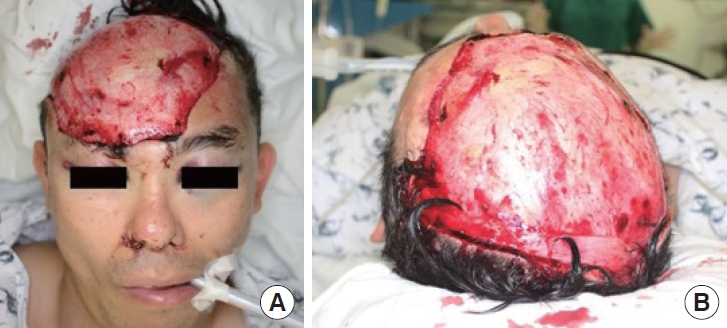
A 44-year-old man with a traumatic scalp avulsion. (A) Frontal view. (B) Top view.
The amputated part was prepared first to reduce the ischemia time. To set the orientation of the avulsed scalp flap, a full-skull rapid prototyping (RP) model was used as an anchorage during shaving and irrigation. Placing the amputated part on the three-dimensional RP model provides a more uniform distribution, in contrast to positioning it on a flat surface. This approach facilitates the evaluation of its orientation and allows for meticulous preparation, ensuring the absence of undesirable wrinkles, which is highly beneficial for subsequent insetting (Fig. 2). Hair was trimmed, followed by the removal of visible dust and debris. The scalp was sterilized using massive irrigation. Inspection of the vascular stump enabled identification of the right superficial temporal vessels under a microscope.
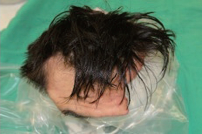
Avulsed scalp before preparation using rapid prototyping model for orientation and sterilization.
The surgery was performed under general anesthesia 10 hours after the trauma. We meticulously dissected approximately 2.0-mm recipient vessels under a microscope to locate the frontal branch of the right temporal vessels. Using an end-to-end anastomosis technique (Fig. 3), we performed microanastomosis on the right superficial temporal artery and vein, resulting in a total ischemic time of approximately 11 hours. In addition, we performed neurorrhaphy on the right supratrochlear and supraorbital nerves. Regrettably, we could not perform neurorrhaphy on the severed nerves beyond the forehead on the opposite side.
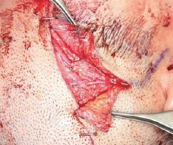
End-to-end anastomosis of the right superficial temporal artery and vein.
The avulsed scalp was anchored using skin sutures. After insetting, the replanted flap showed congestive signs on the frontal area, with a short refilling time of approximately 1 second. Initially, we intended to use the left temporal vein as the recipient for an additional vein anastomosis. However, this proved difficult during the anastomosis because of the considerable separation between the amputated scalp vein and the designated recipient vessel, escalating procedural complexity. After completing a single anastomosis, we checked flap perfusion using indocyanine green (ICG) fluorescence imaging, which provides high sensitivity in detecting well- and non-perfused tissue [6]. Because imaging results showed adequate washout, the procedure was terminated after inserting two negative pressure drainage tubes and one silastic drain beneath the replanted scalp to prevent hematoma (Fig. 4).
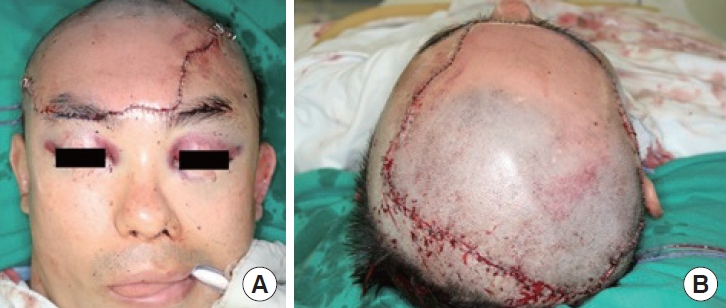
Immediate postoperative photographs. (A) Frontal view. (B) Top view.
Initially, we planned to use heparin for flap survival. However, because of a hemorrhage in the frontal lobe, no anticoagulant or antiplatelet agents were systematically applied during or after the surgery. Blood transfusion was administered until postoperative day 1 with fluid resuscitation management according to hemodynamic parameters. Antibiotics were used to prevent infection up to the day of discharge.
Immediately after the surgery, an evaluation revealed congestion in the flap. However, as we had confirmed adequate washout with intraoperative ICG fluorescence imaging, we considered the likelihood of a vein occlusion to be low and raised the head to 45° early and closely monitored the progress. Three days after the surgery, the flap displayed a refill time of 1.5 seconds and maintained a consistent color. Subsequently, we started removing the stitches, beginning at the facial margin on postoperative day 6 and continuing on postoperative day 14. There were no complications, such as skin necrosis or margin dehiscence.
After 3 months, the patient complained of decreased sensation and sweating on the forehead. However, 6 months after the surgery, all symptoms spontaneously improved, and the forehead sensation recovered without further complaints (Fig. 5).
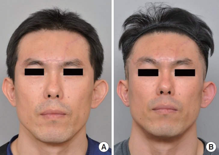
Follow-up photographs at postoperative 3 months (A) and 8 months (B).
DISCUSSION
The vascular supply to the scalp comes from the superficial temporal and occipital arteries. Some reports have suggested that bilateral repair of vessels provides more competent scalp replantation outcomes [3-5,7]. It would be difficult to guarantee that bilateral vessel anastomosis would result in better outcomes, as it is difficult to guarantee this based on the number of vessel anastomoses alone when checking perfusion. However, this case supports the hypothesis that a single artery–vein anastomosis is sufficient for the perfusion of large scalp sections.
The most common cause of replantation failure is insufficient venous drainage, which affects scalp survival [7-9]. In this particular case, we initially attempted to perform an additional vein anastomosis. However, following a thorough evaluation, which included the use of ICG imaging to confirm perfusion and washout adequacy, we determined that flap viability could be confidently predicted without further vein anastomosis. Access to effective tools, such as ICG fluorescence imaging, and new technology to evaluate flap circulation with high sensitivity would help plastic surgeons to predict free flap survival postoperatively and for inoperative decision-making [10].
Apart from the success of anastomosis, ischemia duration is crucial in determining flap survival after replantation; thus, all efforts should aim to decrease the time between trauma and revascularization [4,5,7,8,11]. To facilitate surgical preparation, we meticulously placed the crumpled amputated part on the skull’s RP model. This strategic positioning aimed to flatten the surface and remove any residual hair or foreign objects. Throughout the surgical preparation phase, a diligent approach was maintained to minimize ischemic time by effectively managing the avulsed scalp. This involved promptly orienting and sterilizing the avulsed frontal flap. The surgeon must adhere scrupulously to each step of the scalp replantation procedure, ensuring a seamless and successful progression at every stage.
Microsurgical replantation is the best procedure for esthetic and functional outcomes. After neurorrhaphy, which included supratrochlear and supraorbital nerves, the patient finally showed recovery of sensation and sweating on the scalp 6 months after surgery. Factors such as flap thickness and scarring in the recipient bed may contribute to sensory recovery after free tissue transfer [12]. Without nerve repair, reinnervation appears to occur by nerve ingrowth from the recipient bed, requiring a long time and may induce disorientation [12,13]. Therefore, replantation with neurorrhaphy may result in better functional outcomes than without nerve repair, thus permitting a faster return to a quality of life similar to that before the trauma.
Notes
Conflict of interest
No potential conflict of interest relevant to this article was reported.
Funding
None.
Ethical approval
This case was approved by the Institutional Review Board of Asan Medical Center (IRB No. 2018-0485).
Patient consent
The patient provided written informed consent for the publication and use of his images.
Author contributions
Conceptualization: Woo Shik Jeong. Data curation: Woo Shik Jeong. Writing - original draft: Dongjin Kim, Somin Oh. Writing, review, and editing: Dongjin Kim, Woo Shik Jeong. Supervision: Woo Shik Jeong.
Abbreviations
ICG
indocyanine green
RP
rapid prototyping