Upper lip tie wrapping into the hard palate and anterior premaxilla causing alveolar hypoplasia
Article information
Abstract
Bony anomaly caused by lip tie is not many reported yet. There was a case of upper lip tie wrapping into the anterior premaxilla. We represent a case of severe upper lip tie of limited lip motion, upper lips curling inside, and alveolar hypoplasia. Male patient was born on June 3, 2016. He had a deep philtral sulcus, low vermilion border and deep cupid’s bow of upper lip due to tension of short, stout and very tight frenulum. His upper lip motion was severely restricted in particular lip eversion. There was anterior alveolar hypoplasia with deep sulcus in anterior maxilla. Resection of frenulum cord with Z-plasty was performed at anterior premaxilla and upper lip sulcus. Frenulum was tightly attached to gingiva through gum and into hard palate. Width of frenulum cord was about 1 cm, and length was about 3 cm. He gained upper lip contour including cupid’s bow and normal vermilion border after the surgery. This case is severe upper lip tie showing the premaxillary hypoplasia, abnormal lip motion and contour for child. Although there is mild limitation of feeding with upper lip tie child, early detection and treatment are needed to correct bony growth.
INTRODUCTION
Upper lip tie is uncommon case which makes it difficult for babies to feed breast milk. It is revealed by researches that upper lip tie and tongue tie can make functional problem, but almost no one had bony anomaly caused by lip tie [1-5]. And in most cases, it is not treated if there is no limitation of baby’s growing result for difficulty of breastfeeding [1,2]. There was a case of upper lip tie wrapping into the anterior premaxilla in our clinic. It was tight cord of lip strongly attached from vermilion to premaxilla. We checked up facial three-dimensional computed tomography (3D CT) and found that there was an alveolar hypoplasia caused by severe upper-lip tie. We represent a case of particular upper lip tie showing limited lip motion, upper lips curling inside, and alveolar hypoplasia. It was needed to be released for alveolar growing and lip contour.
CASE REPORT
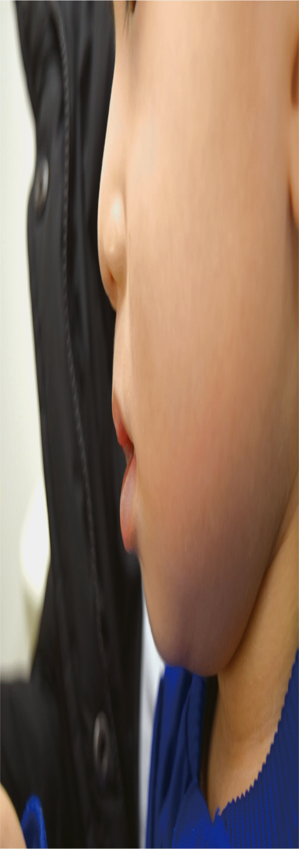
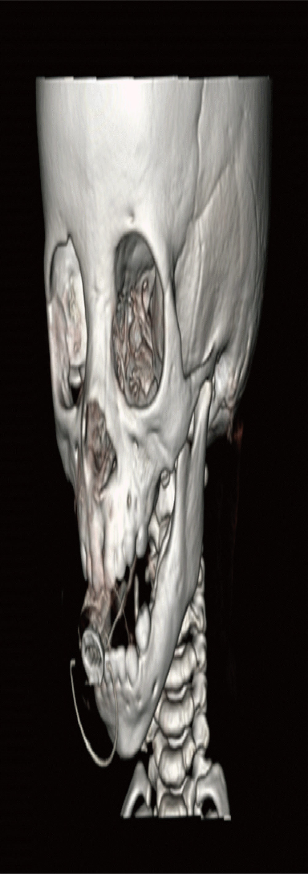
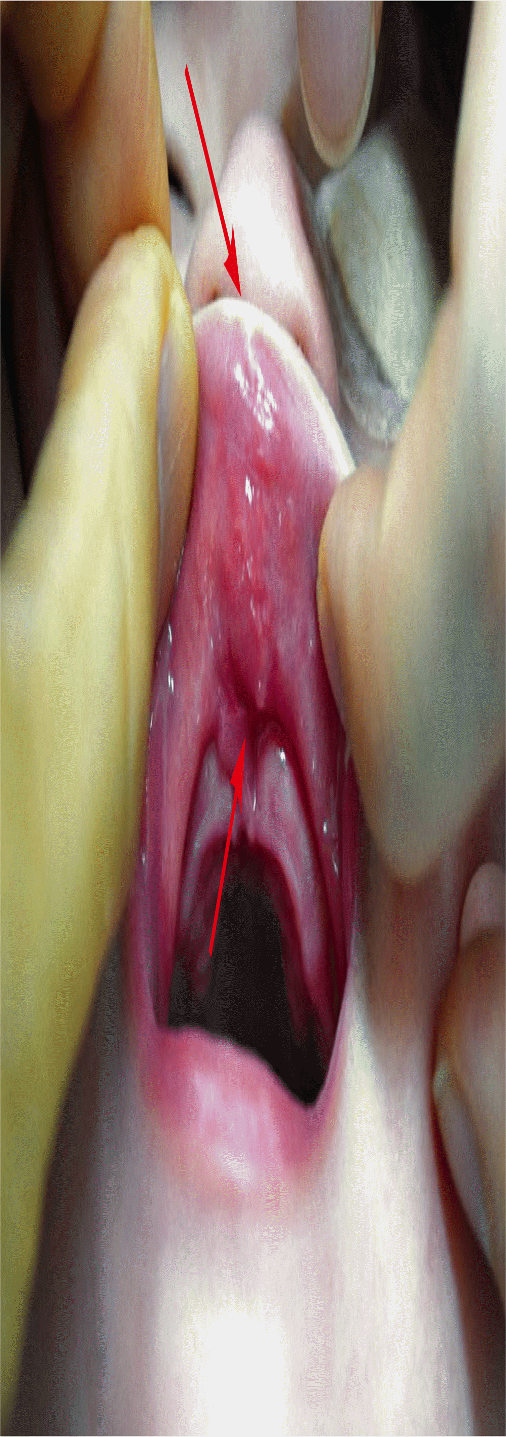
Male patient was born on June 3, 2016 without choromosomal abnormality. He had a deep philtral sulcus, low vermilion border and deep cupid’s bow of upper lip due to tension of short, stout and very tight frenulum. His sucking power was almost normal, but upper lip motion was severely restricted in particular lip eversion from anterior maxilla. We checked up facial 3D CT so that there was anterior alveolar hypoplasia with deep sulcus inside. Because his upper lip tie was toughly attached through hard palate and anterior maxilla, we thought it possibly made alveolar hypoplasia (Fig. 1). We tried to detect any other accompanied congenital malformation, but it was not classified any other congenital disease. We decided to release tie to make him have adequate alveolar and premaxillar growth and lip motion including lip contour. Eight-month-old patient admitted for frenulectomy with Z-plasty of frenulum through upper lip and premaxilla. Resection of frenulum cord with Z-plasty was successfully performed at anterior premaxilla and upper lip sulcus. Frenulum was tightly attached to gingiva through gum and into hard palate. Width of frenulum cord was about 1 cm and length was about 3 cm. Cord was too wide and tough, and it caused the deficiency of alveolus bone between upper incisors, probably causing oral cavity anomaly from childhood (Fig. 2). He gained upper lip contour including cupid’s bow and normal vermilion border after the surgery (Fig. 3). We rechecked facial 3D CT and found that alveolar hypoplasia was almost corrected at postoperative 10 months (Figs. 4, 5).
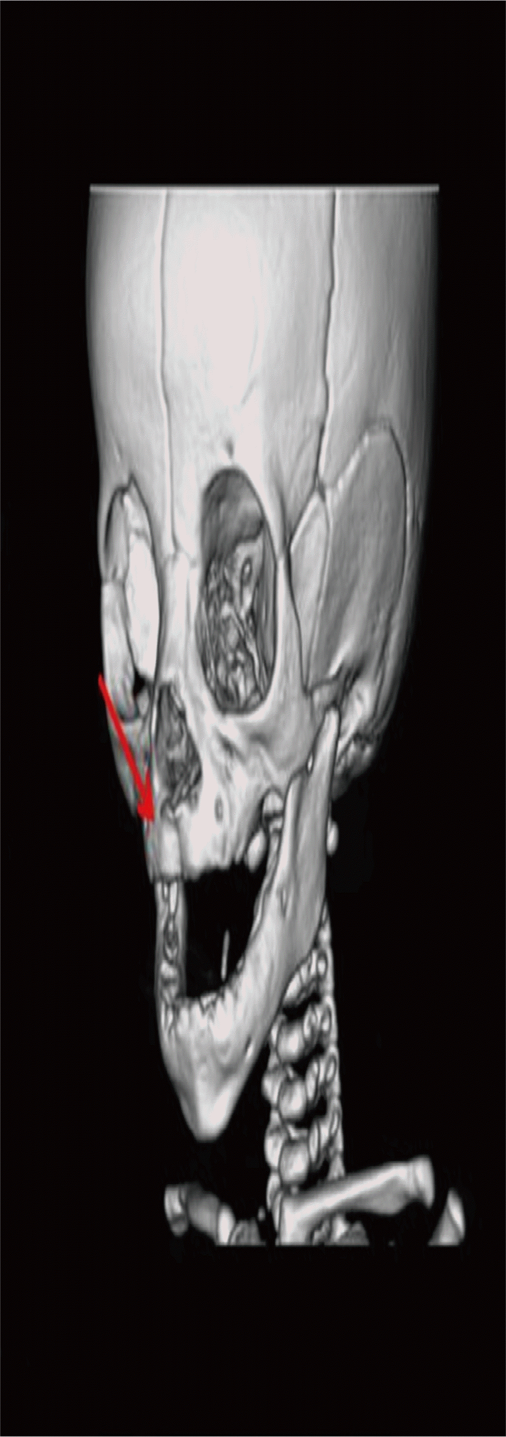
Preoperative facial three-dimensional computed tomography. It shows the bony sulcus of premaxilla and alveolus due to tight, short cord of upper lip frenulum (arrow).
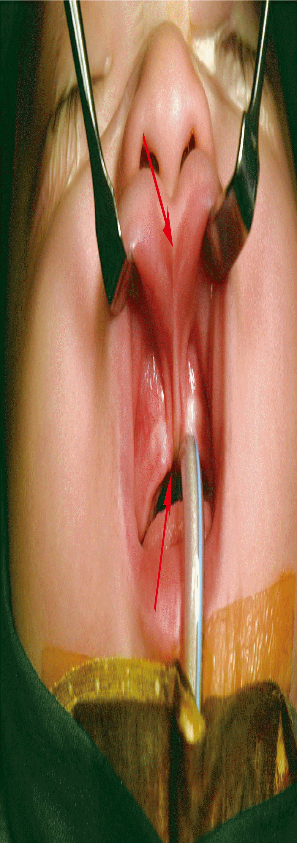
Intraoperative photograph. There was stout frenulum cord wrapped gum into the anterior premaxilla, and invaded into the alveolus. Eversion of lip was not possible due to the cord tightness (arrows).
Postoperative 2 months and 17 days photograph. Cord was resected from premaxilla and gum. Lip was completely released from the alveolus so that upper lip can be everted well after surgery (arrows).
DISCUSSION
Upper lip tie is a benign condition that tends to improve with normal facial growth [6]. It can be contributing factor to breastfeeding difficulty and abnormal lip motion. It may cause ineffective latching but significant functional problem like speech production is not reported yet [1]. Generally, relief surgery for upper lip tie may be indicated when the baby has not enough sucking ability for breast feeding or abnormal lip motion [3]. But there are several cases that upper lip tie alone can cause maxillary diastema, or gap between upper two central teeth [1].
It is reported that frenulotomy of upper lip tie alone results in low recurrence rate and high improvement rate of breast feeding [1,3,7]. For mild upper lip tie, simply dividing of frenulum by iris scissor can be the treatment of choice [7]. But this particular case is severe upper lip tie resulting alveolar hypoplasia, abnormal lip motion and contour for child. And it is very rare and needed to re lease the cord. In this case, upper lip was curling inside because of cord tightness. Thus, we should release inner upper lip by Zplasty after cord resection.
This case demonstrates that tension of upper lip tie itself can make bony hypoplasia and abnormal lip contour for child. Although there is no limitation of feeding with upper lip tie child, early detection and treatment are needed to correct bony growth. Severe case of upper lip tie like in this case, cord resection with releasing by Z-plasty can be successful surgical technique. We should closely observe recurrence and maxillary growth in his growing period.
