INTRODUCTION
Deformities of the external ear canal (EAC) can be caused by tumors, burns, trauma, and malformations. Reconstruction is sometimes required after surgery of the EAC [1,2]. The patency and shape of the EAC must be maintained in order to preserve its functions, including hearing and protection against microbes [2]. Many surgical approaches with varying success rates for EAC reconstruction have been devised, including secondary healing, skin grafting, and a variety of local skin flaps [3].
Among these methods, skin grafting is commonly performed because of its technical simplicity. However, it has been associated with certain complications, including contracture formation, stenosis, and delayed wound healing [3,4]. Proper graft compression and shape maintenance have been suggested as ways to help overcome these shortcomings [5,6]. Many creative ideas have been described in the literature with the goal of providing proper structure and compression to difficult skin graft locations, including quilting sutures, negative-pressure wound therapy (NPWT), foam quilting, and surgical gloves [7-10]. In this report, we describe a case of EAC reconstruction with a skin graft after the resection of a premalignant lesion. The cover of an ear thermometer probe was successfully used as a mold to match the curvature of the EAC.
IDEA
A 55-year-old Asian woman presented to our clinic with a 1-week history of tinnitus in the left ear. The patient denied hearing loss, otalgia, otorrhea, bleeding, or vertigo. No visible lesions were observed in her outer ear. Otoscopy revealed a painless 1.0 ├Ś 1.5 cm patch-like lesion just above the tympanic membrane (Fig. 1). The left tympanic membrane was intact and no hearing impairment was observed on pure-tone audiometry. A further evaluation using computed tomography revealed no involvement of the temporal bone or head and neck structures.
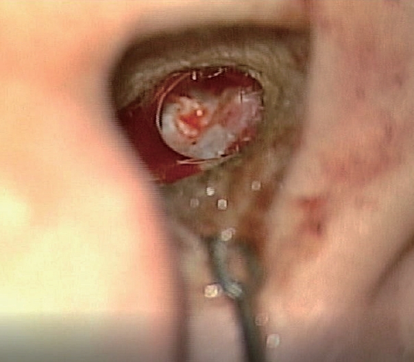
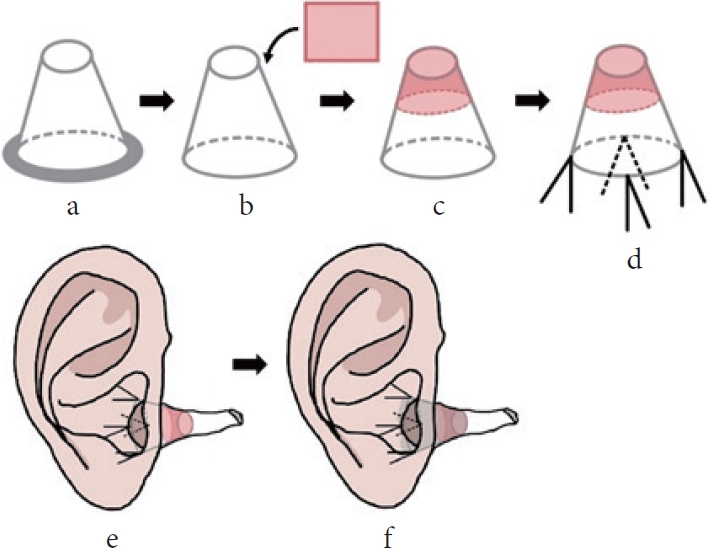
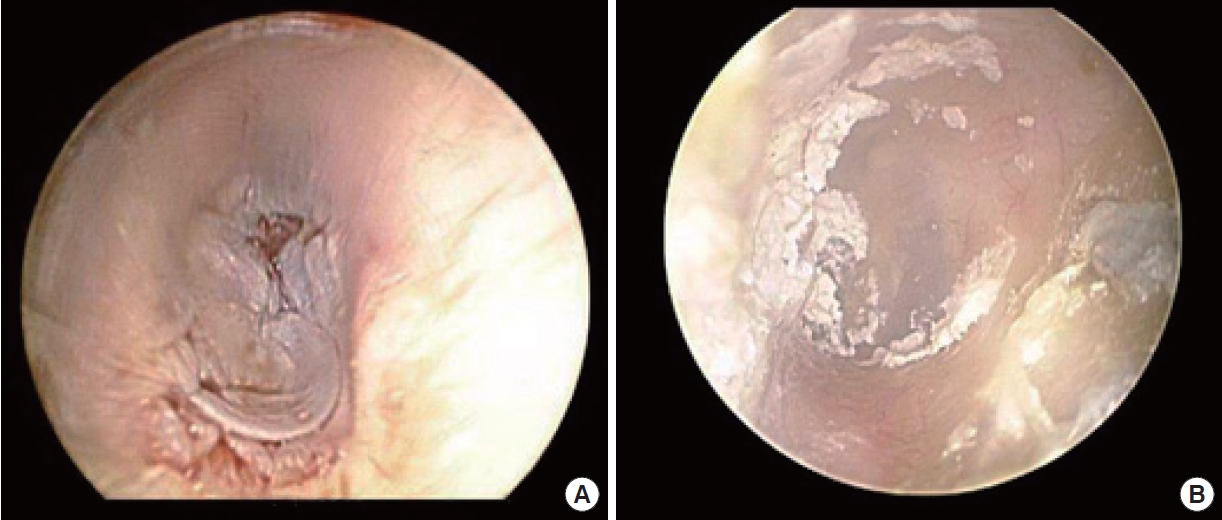
The lesion was diagnosed as actinic keratosis on skin biopsy. Due to the malignant potential of the lesion [11,12], local resection and reconstruction with a split-thickness skin graft (STSG) were planned. The EAC skin was surgically removed with partial cortical bone curettage to preserve the outer half of the EAC and the cranial portion. After resection, the skin defect size was 1.5 ├Ś 2.0 cm (Fig. 2). A 10/1,000-inch layer of skin was harvested with an air dermatome. To maintain the shape of the cone-shaped graft, a sterilized cover of an ear thermometer probe (MC-EP2 for ear thermometer model TH839S; Omron) was used as a mold for the EAC (part a in Fig. 3). This was used to bolster the skin graft on the EAC. In order to place the probe cover deeper, its rim was cut, and the body was punched with a 21-gauge needle to help drain the wound discharge. The harvested skin was placed on the tip of the probe cover and sutured using 5-0 coated Vicryl sutures (Ethicon Inc.) (part c in Fig. 3C). After positioning the skin, a mesh gauze was packed into the cavity of the probe cover and its opening was tagged with 4-0 black silk (AILEE Inc.) (part d in Fig. 3). The probe cover was subsequently placed in the defect using a bolster suture to hold the harvested skin in place and prevent skin graft displacement (Fig. 4). Both the graft dressing and thread gauze used for inner packing were changed every 3 days without disturbing the graft. Polyurethane foam dressing was used to treat the donor site. The wound had healed well without any complications at 2-week (Fig. 5A) and 3-month (Fig. 5B) postoperative follow-up visits.
DISCUSSION
The maintenance of patency during EAC reconstruction is essential for the preservation of hearing function, continuity of access for routine ear cleansing, and physical examinations [2,4]. Various techniques are available for EAC reconstruction that help maintain its patency [3,4]. The three-dimensional curvature of the EAC makes it difficult to perform skin grafting; such curved surfaces require the optimization of techniques [13]. Defects smaller than 2.0 cm may be left to heal by secondary intention or adequately covered with a skin graft. To restore defects larger than 2.0 cm, local flaps and skin grafts may be used, as described in previous research [14]. In our case, a 2.0 ├Ś 1.5 cm defect resulted from the resection of actinic keratosis in the inner portion of the EAC. To minimize donor site morbidity and retain the shape of the canal, we reconstructed the defect using STSG.
Efforts were made to develop a suitable mold, because it is difficult to retain the harvested skin intact against the bony canal [5,6]. Although previous studies have proposed using a nasopharyngeal tube or ear mold stent [5,6], it is difficult to fit a nasopharyngeal tube to match the shape of the ear, and an ear mold stent requires a long manufacturing time. In addition, although ear molds can fit well, their usage period is rather short compared to the cost. NPWT and quilting sutures are also popular methods for skin graft dressing [8,10] that facilitate compression and drainage. They are, however, not appropriate for the narrow space of the EAC and the sensitive tympanic membrane.
Therefore, we used an ear thermometer probe cover as a mold for a skin graft in this case. As it was already made for the ear canal, only minimal additional trimming was required to make it suitable for this purpose. Furthermore, the probe cover facilitated serum imbibition and graft collection without causing canal stenosis or infection. Therefore, the use of a cover of an ear thermometer probe as a mold for an EAC skin graft has the potential to reduce costs and simplify surgery.
To achieve successful wound healing using a skin graft, it is important to maintain the grafted skin in close contact with the wound. Hence, a cover of an ear thermometer probe is a useful graft mold for EAC defect reconstruction using a skin graft.