INTRODUCTION
Solitary fibrous tumor (SFT) is a rare neoplasm, accounting for fewer than 2% of soft tissue tumors. In 1931, it was first reported by Klemperer and Rabin, and SFT is challenging to diagnose and treat. SFT has been referred to using many different terms, including localized fibrous tumor, solitary fibrous mesothelioma, localized mesothelioma, and benign mesothelioma, because its macroscopic and histologic characteristics overlap with those of other soft tissue tumors [1]. However, further research and discovery have made it possible to precisely identify and categorize soft tissue tumors, including SFT. According to the 2017 World Health Organization Classification of Head and Neck Tumors, SFT was categorized as a borderline/low-grade malignancy [2]. It does not have a sex predilection and mainly affects adults in the fifth and sixth decades of life [3]. To date, no environmental risk factors have been associated with SFT risk. SFT is a tumor of mesenchymal origin that mainly develops in the pleura [4]. Roughly 30% of SFTs are found in the extremities, thyroid gland, and vulva, and 6% are observed in the head and neck region [5]. The orbit and the sinonasal tract are the most common sites in the head and neck region, followed by the salivary glands and oral cavity [6]. A literature search of PubMed and Google Scholar on March 31, 2023, using the terms “temporal region,” “temporalis muscle,” “cancer,” “tumor,” “solitary,” “fibrous,” and “mesothelioma” without date or language restrictions yielded only one report of SFT in the temporal region. However, it was a case of capsulized SFT in the sub-dermal layer of the temporal region [7]. This report presents a case of SFT that occurred in the right temporalis muscle under the zygomatic arch in a middle-aged man.
CASE REPORT
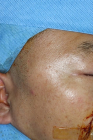
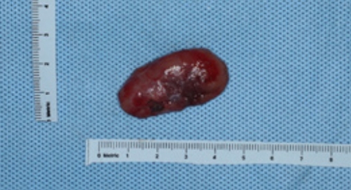
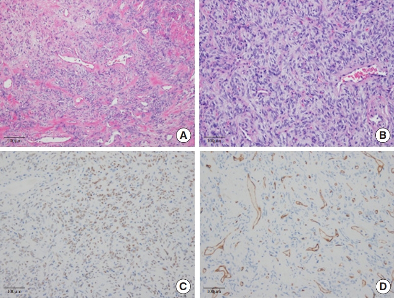
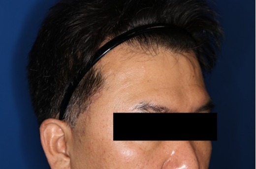
A 47-year-old man with a palpable mass in the right temporal area that had developed 7 months ago, was seen at our outpatient clinic. He complained about the size of the mass, which had gradually increased (Fig. 1). Computed tomography showed a 4.0×2.9×1.4 cm well-defined heterogeneously enhancing mass as a vascular tumor in the right temporalis muscle under the zygomatic arch (Fig. 2). No specific findings were observed in the paranasal sinus, pharynx, and salivary glands. There were also no significantly enlarged lymph nodes in the neck. The patient had no specific past medical history. Under general anesthesia, a hairline incision was performed to avoid neurovascular damage around the temple area and minimize the scar. After elevating the deep temporal fascia, it was confirmed that the mass was inside the right temporalis muscle. The mass was removed from the right temporalis muscle, and the remaining muscle was repaired (Fig. 3). Based on a histopathological examination of the mass, which showed spindle cell proliferation with variable cellularity depending on the area, and immunohistochemical staining of the mass, which demonstrated immunoreactivity for signal transducer and activator of transcription 6 (STAT6), the mass was determined to be an SFT (Fig. 4). According to the three-variable and modified four-variable risk models for the prediction of metastatic risk, the metastatic risk of this tumor was low. The patient was discharged without any complications and was followed up regularly. No recurrence or metastasis was observed for 3 months (Fig. 5).
LITERATURE REVIEW
We conducted a literature review of intramuscular SFT occurring in the head and neck region, analyzing articles available on PubMed and Google Scholar up to 2023. We gathered data on the age, sex, tumor size, location, recurrence trends, treatment, immunohistochemistry, outcome, and follow-up period.
In total, five cases were analyzed, including the one presented in this study (Table 1) [8-11]. Of these five cases, two involved males, two involved females, and one did not specify the patient’s sex. The average age at diagnosis was 48.4 years, with a range from 27 to 75 years. Tumor measurements were based on the longest axis, ranging from 1.5 to 5 cm, with an average size of 3.3 cm. One case did not provide information about the tumor size. The tumors were located in various locations, including the right lateral and medial rectus muscles, the left lateral rectus muscle, the left masseter muscle, the left sternocleidomastoid, and the right temporalis muscle.
At the time of the hospital visit, three patients reported experiencing painless swelling. Those who complained about intraocular SFT involving the rectus muscle reported ocular symptoms. One patient experienced a progressive limitation of extraocular motility and worsening exophthalmos, while another patient had a history of proptosis and chemosis.
SPT has generally nonspecific features on radiography. However, computed tomography tends to show a mass that is welldefined, sometimes isodense to skeletal muscle, and occasionally lobulated, with heterogeneous contrast enhancement that results from the extensive tumor vasculature. T1-weighted magnetic resonance imaging (MRI) presents intermediate intensity, while T2-weighted MRI demonstrates and variable hypointensity to hyperintensity, which reflects to fibrous and cellular or mycoid regions [2]. In four cases, contrast-enhanced T1-weighted MRI showed an enhanced mass involving muscles [8-11]. In one case, there was a well-defined heterogeneously strong enhancing mass in the right temporalis muscle with vascularity.
Information regarding immunohistochemistry was provided for four cases (Table 1) [8,9,11]. All cases demonstrated immunoreactivity for CD99 and were negative for S-100. Three of the cases tested positive for CD34, while one tested negative. B-cell lymphoma 2 (Bcl-2) immunoreactivity was reported in three cases. One case showed immunoreactivity for desmin, while two cases tested negative.
Only one case report provided information on the molecular characterization, which revealed the absence of COL1A1-PDGFB fusion transcripts. Of the five cases reported, four involved primary SFT, while one case was a metastatic SFT originating from a pleural SFT.
All cases were initially managed with surgical excision, with some requiring additional treatment. There was one case where the treatment was not reported. Another case involved radiation therapy, chemotherapy, and exenteration of the orbit due to local recurrence and malignant transformation. A case of metastatic SFT, originating from pleural SFT, was treated with radiation therapy.
There are so few known cases of intramuscular SFT developing on the head and neck that its prognostic outcomes cannot be predicted in a generalizable sense. However, cases involving recurrence or metastasis have shown a poor prognosis. In our study, the patient did not exhibit tumor recurrence or complications, including functional impairment, 8 months post-surgery. Therefore, additional research and a larger case study are necessary to establish a robust, evidence-based guideline for treating intramuscular SFT in the head and neck.
DISCUSSION
SFT is a fibroblastic tumor with prominent vasculature showing branches, thin walls, and dilatation, and cases in the head and neck region mainly develop in the oral and nasal cavities [6]. Most SFTs present as painless, slow-growing masses [6]. The diagnostic criteria of SFT are a mass showing spindle to ovoid cells arranged around a branching and hyalinized vasculature on histopathology, variable stromal collagen deposition on cytology and CD34 and/or STAT6 expression on immunohistochemistry [6].
Several markers that show positivity in SFT are CD34, CD99, Bcl-2, and STAT6. Some markers are also consistently negative in SFT depending on the location, such as endothelial membrane antigen (EMA) and S100 [12]. The CD34 antigen is a transmembrane cell surface glycoprotein (110 kDa) that is observed on myeloid progenitor cells. Spindle-shaped neoplastic cells are immunoreactive to CD34. Thus, several tumors, such as SF, gastrointestinal stromal tumor, epithelioid sarcoma, Kaposi sarcoma, dermatofibrosarcoma protuberans, can show CD34 positivity on immunochemical staining [13]. The CD34 antigen has been found in 95% to 100% of cases of SFT [12]. In the past, it was considered the most consistent marker of SFT. However, it lacks specificity for the exact identification of SFT from other tumors. The recent discovery of the NGFI-A binding protein 2 (NAB2)-STAT6 fusion gene has led to a more precise diagnosis of SFT. NAB2-STAT6 fusion is specific for SFT, and the demonstration of the fusion gene can be helpful in challenging cases [12]. However, the molecular tests are expensive and are not available at every center. Thus, STAT6 expression has been utilized as a proxy for detecting the fusion gene. STAT6 belongs to the STAT protein family, which contains seven members, is closely linked to interleukin-4 and interleukin-13 signaling, and plays an important role in TH2 polarization in the immune system [14]. Strong STAT6 immunoreactivity has been confirmed to be highly sensitive and specific for SFT [12]. The immunohistochemical stains of the mass in this patient showed immunoreactivity for STAT6, CD99, Bcl-2, and focal positivity for actin. Desmin, S-100, EMA, and CD34 were unreactive.
The histology of SFT shows haphazardly arranged spindled to ovoid cells with pale and indistinct eosinophilic cytoplasmic regions within a variable collagenous stroma deposition admixed with branching and hyalinized staghorn-shaped bold vessels [6]. Microscopically, the patient’s tumor revealed short spindle cell proliferation with variable cellularity depending on the area. The hypocellular area showed spindle cell proliferation admixed with branching and hyalinized blood vessels and stromal collagen. The hypercellular area revealed closely spaced short spindle cells with no intervening stroma (Fig. 4).
SFT is generally considered to be a benign tumor, but several cases of metastasized SFTs have been reported [15]. SFT cases that show malignant histologic features, such as areas of necrosis, high cellularity, large tumor size (>10 cm), atypical mitotic figures, and high mitotic count (>4 per 10 high power fields) are more likely to recur and metastasize [16]. No significant difference has been shown between benign and malignant SFTs in terms of age, sex, and tumor site [17]. The patient was under 55 years of age and showed a low mitotic count of 0−1 mitoses/mm2. In addition, since the tumor size was <5 cm and tumor necrosis was not observed, according to the three-variable and modified four-variable risk models for the prediction of metastatic risk, this tumor was low-risk (0–1 point).
For localized SFT, surgery is the treatment of choice. After complete surgical resection with clear margins, the 10-year survival rates are between 54% and 89%. Radiation therapy can be used to improve local control, but it does not affect overall survival. Chemotherapy is often used as a final effort, but many patients do not respond [18]. Targeted treatments, such as angiogenesis inhibitors, are likely to be effective in managing disease [19]. In this patient, the SFT was successfully removed from the right temporal muscle by a surgical procedure, and no recurrence or adverse effects were observed during a 3-month follow-up. Since the metastatic risk was low, the patient did not undergo radiation therapy and chemotherapy.
The attachment of the temporalis muscle is located superior to the zygomatic arch, above the fascia and bone in the temporal fossa, and inferior to the coronoid process of the mandible and along the mandibular ramus. The temporalis muscle is supplied by anterior deep temporal artery in anterior area, posterior deep temporal artery in middle area and middle temporal artery in posterior area [20]. The major function of the temporalis muscles is to close the jaw, while the middle fibers are responsible for retrusion of the mandible bilaterally. When the temporalis muscle acts unilaterally, it causes the mandible to deviate toward the same side [21]. Injury of the temporalis muscle during surgery often leads to postoperative atrophy, potentially resulting in severe asymmetry. The causes of asymmetry include atrophy of the distally transected muscle or the entire muscle, as well as incorrect positioning of the muscle during the completion of surgery [18]. After surgical resection of the SFT in the temporalis muscle, the patient did not report discomfort during mastication or cosmetic dissatisfaction.
Soft tissue tumors in the temporal region occur infrequently. The following conditions should be included in the differential diagnosis: temporal muscle herniation through a defect of the anterior temporalis fascia, arteriovenous malformations, temporal arteritis, myositis ossificans, metastasis, soft tissue sarcoma, soft tissue osteoma, epidermoid cyst [22,23]. SFT is a rarely occurring tumor in the temporal region, and no case of SFT in temporalis muscle has previously been reported. Although SFT in the temporalis muscle is rare, surgeons must consider the possibility of SFT in the differential diagnosis of patients with a mass in the temporal region.